Lupine Publishers | Journal of Surgery & Case Studies
Introduction: Rib fractures are a common injury after road
traffic accidents. While most simple rib fractures heal well, multiple
rib fractures may result in acute life-threatening complications or
chronic disability and work loss. Though surgical fixation of rib
fractures has most commonly been restricted to multiple rib fractures
with flail chest, there has been a recent interest in fixation of
multiple rib fractures with chest deformity to preclude chronic
disability and loss of work.
Case Report: We report the case of a 34 year male with multiple rib fracture and chest deformity due to multiple, displaced fractures of 3rd to 10th ribs on the left side. He was treated with open reduction and internation fixation of ribs with 2.4mm titanium reconstruction plates and screws. The emerging indications of rib fracture fixation, as seen in this patient, are discussed.
Conclusion: Longer duration of hospital stays and delay in returning to normal life result in poor quality of life and add to direct and indirect treatment expenses. A case-based approach is essential in the decision-making for surgical fixation of multiple displaced rib fractures.
Keywords: Rib Fractures; Fracture Fixation; Chest Deformity
Rib fractures are one of the most common injuries after
road traffic accidents. Most simple rib fractures heal well with
minimum intervention. But multiple rib fractures may require use
of mechanical ventilation and sometimes surgical management
[1]. Thoracic trauma comprises 10-15 % of all trauma and are the
causes of death in 25 % of all fatalities due to trauma [2]. We present
a case of multiple rib fractures and chest deformity and present the
outcome of surgical fixation and its significance.
A 34 year male, a bus conductor, was brought to our hospital in
the emergency room with an alleged history of road traffic accident.
He sustained mild head injury with a history of loss of consciousness
and there were multiple abrasions all over his body. He complained
of severe excruciating pain during breathing and movements of
left arm, with a pain score in VAS scale at 8-9(0-10). Pain was nonresponsive
to analgesics. He had significant depression of the chest
wall on the left side; chest wall movements were equal bilaterally.
Computed tomography of the brain showed no parenchymal injury.
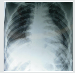
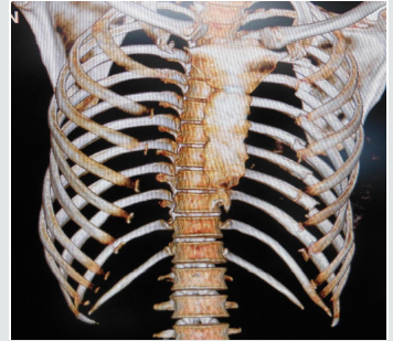
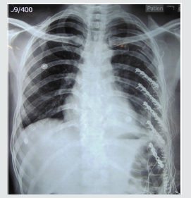
Plain chest radiograph (Figure 1) and computed tomography with
3D reconstruction (Figure 2) demonstrated multiple, displaced
fractures of 3rd to 10th ribs on the left side. There was no evidence of
pneumothorax, hemothorax or lung contusional injury.
There were no signs of pleural tear after fixation, as clinically confirmed by positive pressure ventilation. The wound was closed in layers with a vacuum drain in-situ. He made a rapid recovery with marked reduction in his pain and discomfort (VAS score of 5) on post-operative day 1. The chest wall deformity was fully corrected. He was discharged on the 3rd post-operative day. Patient was last followed-up at 7months. The fractures had united (Figure 5) and recovery was uneventful. He had returned to work 3weeks following surgery.
Incidence of rib fracture reported by various studies ranges
between 7 - 40 %. Most commonly 4th - 9th ribs are fractured.
Fractures of upper ribs (1st & 2nd) usually signify severe trauma with
increased risk of great vessel injuries [2]. Recently there has been a
resurgence of interest in the surgical management of rib fractures
[3,4]. Indications for surgical fixation of rib fractures include flail
chest, severe chest wall deformity, failure to wean from mechanical
ventilation, chronic pain or disability, pulmonary herniation, nonunion
and “on the way out” after thoracotomy [5]. Initial research
suggests that in select patients, operative management of chest
wall injuries is a promising treatment option. Granetzy et al. [4] in
2005 randomised 40 patients who experienced fractures of 3 or
more ribs to receive either conservative or surgical treatment and
the results showed that patients in the surgical group experienced
significantly fewer days on mechanical ventilation, decreased stay
in the Intensive Care Unit and hospital stay and less restrictive
pattern on pulmonary function tests 2 months after treatment [6].
Similar results were found by Nirula et al. [5] in 2006 where they
treated 60 patients with rib fractures [7]. Favourable long term
outcomes of patients undergoing surgical chest wall stabilization
was documented from a prospective study by Lardinois et al. [8],
who had done surgical stabilization of 60 patients of chest wall
injuries from 1990-1999.
Rib fractures have been associated with significant disability and loss of work [9]. Hence selected patients with multiple rib fractures but without flail chest have been hypothesized to benefit better from open reduction with internal fixation than from nonoperative treatment [10,11]. All existing surgical indications are relative. Surgical repair has been attributed to possible sooner return to work and usual activities [5,12]. In a retrospective study by Solberg et al on 16 patients of unilateral rib fracture and chest wall deformity, the overall recovery of the surgically treated patient was much earlier than that of those who were treated conservatively [13]. However, no cohort study is available to confirm the beneficial effects of surgical fixation for multiple rib fractures without flail chest [5,12]. Treatment must be individualized on the basis of the patient’s fracture pattern, overall medical condition, and functional status [12]. This patient presents an ideal scenario where a surgical fixation of the rib fracture would result in better clinical outcomes and reduce the morbidity of prolonged pain and disability and loss of work.
The most preferred modality of treatment of rib fractures is
non-operative, with analgesics and active chest physiotherapy.
However recovery is prolonged or associated with complications,
especially in the presence of multiple rib fracture, floating ribs or a
flail chest. Longer duration of hospital stay and delay in returning
to normal life also result in poor quality of life and add to direct and
indirect treatment expenses. Hence, it is rational to manage certain
patients with multiple rib fracture surgically to reduce morbidy,
mortality and loss of work. Clinical message: The report stresses
the need to make a case-based approach in decision-making and
the need to have a lower threshold for surgical fixation in the
presence of multiple displaced rib fractures. Further cohort studies
are needed to confirm the benefits of internal fixation of multiple
rib fractures in the absence of flail chest.
Abstract
Case Report: We report the case of a 34 year male with multiple rib fracture and chest deformity due to multiple, displaced fractures of 3rd to 10th ribs on the left side. He was treated with open reduction and internation fixation of ribs with 2.4mm titanium reconstruction plates and screws. The emerging indications of rib fracture fixation, as seen in this patient, are discussed.
Conclusion: Longer duration of hospital stays and delay in returning to normal life result in poor quality of life and add to direct and indirect treatment expenses. A case-based approach is essential in the decision-making for surgical fixation of multiple displaced rib fractures.
Keywords: Rib Fractures; Fracture Fixation; Chest Deformity
Introduction
Case Summary
Figure 3: Intraoperative picture demonstrating placement
of 2.4 mm titanium reconstruction plates and screws to fix
the fractures.
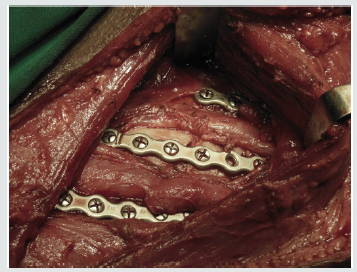
Considering the presence of chest wall deformity and multiple
consecutive rib fractures, surgical stabilization of the ribs was
planned. Under general anesthesia, patient was positioned on left
lateral position, and through a single lazy- S incision starting from
lower border of scapula with a length of 6 cm, lattisimus dorsi
muscle was exposed and split along the fibers and access to the ribs
was made by stripping off the intercostal muscles. The 6th to 10th
ribs were reduced and fixed with 2.4 mm titanium reconstruction
plates and screws (Figures 3 & 4).
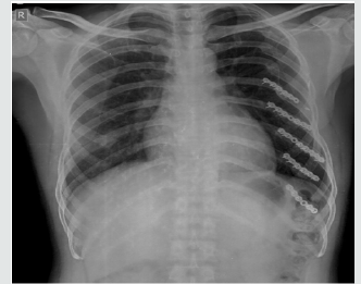
There were no signs of pleural tear after fixation, as clinically confirmed by positive pressure ventilation. The wound was closed in layers with a vacuum drain in-situ. He made a rapid recovery with marked reduction in his pain and discomfort (VAS score of 5) on post-operative day 1. The chest wall deformity was fully corrected. He was discharged on the 3rd post-operative day. Patient was last followed-up at 7months. The fractures had united (Figure 5) and recovery was uneventful. He had returned to work 3weeks following surgery.
Figure 5: Anteroposterior chest radiograph at 7 months
following surgery showing fracture consolidation.

Discussion
Rib fractures have been associated with significant disability and loss of work [9]. Hence selected patients with multiple rib fractures but without flail chest have been hypothesized to benefit better from open reduction with internal fixation than from nonoperative treatment [10,11]. All existing surgical indications are relative. Surgical repair has been attributed to possible sooner return to work and usual activities [5,12]. In a retrospective study by Solberg et al on 16 patients of unilateral rib fracture and chest wall deformity, the overall recovery of the surgically treated patient was much earlier than that of those who were treated conservatively [13]. However, no cohort study is available to confirm the beneficial effects of surgical fixation for multiple rib fractures without flail chest [5,12]. Treatment must be individualized on the basis of the patient’s fracture pattern, overall medical condition, and functional status [12]. This patient presents an ideal scenario where a surgical fixation of the rib fracture would result in better clinical outcomes and reduce the morbidity of prolonged pain and disability and loss of work.
Conclusion
For more Lupine Publishers Open Access Journals Please visit our website:
For more Surgery Journal articles Please Click Here:
To Know More About Open Access Publishers Please Click on Lupine Publishers

No comments:
Post a Comment