Showing posts with label Journal of Surgery. Show all posts

Friday, February 25, 2022

Lupine Publishers | Primary-Simultane Operation Pri Politrauma

 Lupine Publishers | Primary-Simultane Operation Pri Politrauma


Abstract

The author examines the current problems of modern medicine associated with the growth of injuries. In recent decades, not only has the number of injuries increased, but so does the severity of the damage. There were many problems, both in the organization of treatment of victims, and in the methods of surgical correction of injuries, especially if they are combined - multiple, and at the same time injured up to a dozen or even more persons. This is most often the case in road traffic accidents, and they sometimes occur on sections of highways, away from the regional and regional centers, where the main medical staff are concentrated, and in rural areas, where hospitals operate with very limited medical staff. For example, the staffing of the CRB usually provides surgical and trauma departments for 40-60 beds, that is, assistance is planned. or a surgeon, or a traumatologists, but often come persons with trauma of those organs and systems that are to be treated by a neurosurgeon, angiosurgeon and other specialists. The difficulty lies not only in the absence of them, but also in the tools for such operations. One doctor of the CRB receives a load, which in the regional center is allowed 5-10 or more specialists of surgical profile. In this situation, doctors of rural hospitals should have the technique to perform surgery in any form of trauma, but for this they must pass the school of training in leading surgical centers, and now they are mostly former students who know everything, but nothing they know how to do it. Admittedly, the increase in fatalities from injuries is partly due to this factor. The author gives an instructive observation of polytrauma, which fits into the context of the problem at hand.

Keywords: Polytrauma; Surgical treatment; Treatment

The Aim of the Study

To show the importance of the surgeon’s training in providing emergency care to victims of polytrauma

Introduction

Currently, there is a widespread increase in injuries, which is due to the rapid mechanization of work and recreation of the person. With the increase in injuries, there were many problems in the organization of treatment of victims with different types of injuries. The role of peripheral treatment facilities (mainly CRB) in providing effective assistance to traumatized persons has also increased, taking into account the mass migration of the population during the warmer months from cities to villages [1]. The structure of injuries has also changed significantly - the number of combined, multiple and combined multiple injuries is increasing. At the same time, the combined injury includes damage to different systems, and to multiple - the same [2]. With the one-time increase in the number of injured organs and systems, attitudes on surgical aggression have changed, and this has been greatly facilitated by recent advances in anesthesiology, allowing the most severe and pro-longed operations [3,4]. Although until very recently it was thought that expanding the volume of surgery on the human body is dangerous for his life [5,6]. However, the healing process of victims with polytrauma, dictates the need not only to own, but also to perform complex simulative manipulations on various organs and systems. In primary osteosynthesis performed in a combined injury, poor treatment results are twice as common as in other methods of cross-section, and the consolidation of the fracture occurs on average 1 month faster [7-9]. X-ray diagnosis is also needed to clarify the nature and type of injury, but it increases the radial effects on humans [10]. For this reason, it is necessary to resort to magnetic resonance imaging (MRI), as well as to be able to perform ancillary manipulations, including laparocenteza, in case of suspected abdominal injuries [11,12]. However, the leading role in the diagnosis of all injuries still belongs to clinical methods of examination, and they future doctors master during their studies at the university. On this basis, it is necessary to strengthen control over the quality of training of future health professionals in order to eliminate the appearance of ballast, which only interferes with the goal - reducing fatality from injuries, if in reducing their number they are powerless, for it is a national problem.

Material and Methods

In one of the district hospitals of Krasnodar region observed 59 victims with combined-multiple trauma, of which 41 (69%) were affected. primary simulates were performed (from 2 to 6). The rest had 18 (31%) Victims and this was due to their extremely severe condition, or because of the fracture of the bones of the base of the skull, or because of a closed heart injury, limited only to conservative methods of treatment - skeletal stretching, skeletal hanging of the lower limb. All of these patients died five to 18 hours after hospitalization. All patients (41) who had surgery, had multiple fractures of tubular bones, 32 - traumatic brain injury, 6 - closed rupture of abdominal organs, 3 - a fracture of the spine, 3 - pelvic bones, 12 - ribs, 10 – bones. A total of 204 organs and tissues were injured, ranging from 2 to 8 in one person. At the time of hospitalization, everyone had a second- to third-degree shock. The patients were between 13 and 52 years old. The diagnosis was based on clinical, X-ray, endoscopic, instrumental and laboratory examination. All of these patients had metal osteosynthesis of tubular bones (from 2 to 5, only 86), 4 - removed spleen, 2 - stitches on the liver, 6 - trepanation of the skull, etc. Of the 41 victims, 19 had simulated operations carried out 2-6 hours after hospitalization, and the rest within 1 day. The duration of surgical aggression ranged from 2 to 6 hours. Operations were performed under general end tracheal anesthesia with the use of controlled breathing hardware. Two traumatologists took part in the surgical intervention with or without the involvement of a surgeon. The 23-year-old man was admitted after a road accident in a state of extreme severity - pulse filamentous, AD - is not determined, breathing superficial 6-8 in 1 minute. On examination - on the face a lot of abrasions, a parting squint, pupils slightly enlarged and sluggishly react to light, in the left temporal area of the hematoma, from the left ear canal released a blood-bracing liquor (on the X-ray revealed a fracture of the temporal bones). The victim has a closed fracture of both forearms and lower third of the left thigh, and an open middle third of both shins and the middle third of the right thigh. The man was intoxicated. The trachea intubation was carried out and superficial anesthesia was carried out against the background of hardware breathing. After 6-8 hours, the blood pressure rose to 100/50 mm hg. pillar, but soon began to fall, and the stomach to increase in volume. Performed laparocenteza, which revealed blood in the abdominal cavity. Suspected two-moment rupture of the spleen and on vital signs performed laparotomy, which confirmed the diagnosis. A splenectomy was performed, and simultaneous operations were performed on the bone and joint system with the involvement of two more hospital traumatologists, i.e. two teams of specialists worked simultaneously. Sustained metorosemetalostheism of both tibia bones and on Rush of both femurs was performed. The simultaneous surgical creativity lasted about 6 hours, with 5 operations performed. Closed reposition of fragments of both forearms with plaster bandage. The general superficial anesthesia lasted about 2 days, and all this time the hardware breath was carried out. The hospital treatment lasted almost 3 months. There were no complications after the surgery. Gradually, all the fractures consolidated. However, he could not walk without crutches because of pain in his left hip joint. The resulting radiation load did not allow X-rays of this joint, and it was sent to the regional center for MRI. Magnetic resonance imaging unexpectedly revealed an old medial fracture of the cervix of the hip, which was the cause of pain and dysfunction of this joint. All previously diagnosed fractures have been consolidated. He refused hip neck surgery. For 1 year he was disabled in the 2nd group. Further communication with him was lost.

The Result

All the victims safely underwent surgery, but 2 (4.8%) after 3-5 days developed a fat embolism of the cerebral vessels, from which they died. Thus, out of 59 patients with polytrauma, only 20 (33.9%) died. Thirty-nine survivors had no early post-operative surgery. The duration of inpatient treatment in them on average was 45 days (36 to 90 days). The duration of incapacity in 5 patients was 6 months, 21 - 8 months, 10 - more than a year. 3 (7.7%) patients had a persistent disability - 2 it was caused by the consequences of a severe traumatic brain injury, and 1 - a false joint of the cervix of the hip.

Discussion

Thus, the performance of primary Simultane operations in combined-multiple trauma is practically and economically justified activity. They make it possible to successfully combat shock by creating stability of fragments and early activation of victims. The very method of operation, which allows to use existing metal structures effectively, is of great importance in the outcome of the operation. The materials presented in the article fully confirm the data of the literature that these operations reduce the number of postoperative complications by 2 times; on average for 1 month accelerate the consolidation of breaks, which accelerates the rehabilitation of victims. The cause of failure sits largely depends on the severity of the brain and heart injury, and this problem is now waiting to be solved.

Conclusion

Primary simultane operations in combined-multiple trauma can be attributed to the achievements of modern medicine. They allow doctors in polytrauma to actively influence the correction of homeostasis disorders and accelerate the consolidation of bone fractures, which has a clear economic effect. The introduction of new surgical technologies will lead to further progress in the treatment of polytrauma.

Read More Lupine Publishers Surgery and Case Reports Journal Articles: https://surgery-casestudies-lupine-publishers.blogspot.com/.


Friday, December 3, 2021

Lupine Publishers | Multidisciplinary Management of Elderly Cancer Patients: The Radiation Oncologist’s Point of View

 Lupine Publishers | Journal of Surgery and Case Studies


Abstract

Oncological treatments of elderly patients are extremely complex; so far there’s no agreement even on the definition of “geriatric patient”. From the point of view of global health, the problem is of the outmost importance as the number of older patients will increase dramatically in the next years, leading to a change in world epidemiology with a significant increase of chronic-degenerative diseases such as cancer. For this reason, it’s mandatory to provide clinical oncologists with multidisciplinary algorithms aiming at the best treatment of older cancer patients.

Materials and Methods

The complexity of oncological treatments for elderly patients starts from the very beginning, since the definition of “geriatric patient” is not univocal. From the point of view of public health, the problem is of the outmost importance. since nowadays there are 600 million people over 65, in 10 years their number will overcome 1 billion and in 2050 there will be nearly half billion people over 80 [1-3]. Such an increase of longevity will lead to a change in world epidemiology with a significant increase of non-communicable disease such as cancer. In 2030 the annual incidence of new cases of cancer in aged people will be 13,7 million and, which is even more important, nearly half of such cases will be in low-income countries [4]. Very few clinical trials (which are the cornerstone of Oncology) take older patient into account, and usually they are very selected cases [5-8], quite differently from every day’s practice, so there’s paucity of data about care of older patients, which makes clinical oncologist’s task even harder [5].Moreover, at least till few years ago, most guidelines and recommendations only consider chronological age in order to determinate the treatment’s choice, and this policy has led to over- or under-treatments [5]. With the purpose of optimizing the treatment’s choice it is imperative to focus on the concept of biological or functional age, in opposition to merely chronological age [6,7].

We should briefly review the physiological modifications caused by aging, which affects every organ and apparatus. Considering for instance Nervous System, there is a decrease of cortical volume and of synaptic density, which leads to a weaker memory and attention. Cardiovascular system is strongly affected, with a diminished cardiac output, increased arterial stiffness, slower modulation of cardiac frequency etc. [9]. Osteo-muscular apparatus is involved too, with a decrease of bone density leading to an increased risk of fracture and a sarchopenia which causes decreased physical activity with parallel increased fatigue and asthenia [10]. Additionally, for most of oncologic treatments, liver and kidney’s function is crucial; with their reduction, drug toxicity increases. In some aged patients an aforementioned change is plain; in other they can be silent in conditions of balance, becoming evident in stress situations such as a malignant disease and its treatments [1].

Biomolecular Markers of Aging

Aging is an extremely complex phenomenon, showing deep differences among individuals, consequently so far, it’s difficult to identify biological markers which enable us to divide subjects of the same chronological age into different functional ages. Several markers have been suggested, starting with markers of systemic inflammation such as CRP, D-dimer, IL 6 [8]. They are easily quantifiable, and they’ve been associated with functional decline in aged people, but their levels are influenced also by frankly pathological conditions like infections and cancer itself [11,12]. Markers of cellular aging have been considered too, such as telomeres or cell cycle components [8]. Dosing such markers is anyway extremely expensive, and moreover they have a significant interindividual variability. Another marker which could document a link between cancer and aging is P16 INK 4A, which has been showed to increase in aged breast cancer patients receiving chemotherapy [13]. Nevertheless, all of these markers are, so far, not completely validated and reliable.

Geriatric Assessment

As long as validated and reliable biomarkers are not established, the best way to assess a geriatric cancer patient is clinical evaluation [14-17]. Geriatric assessment is a multidisciplinary and multi parametric evaluation which takes into account physical aspect, nutritional status, neurological and cognitive status and even social support [18,19].

Comorbidities

When planning an oncological treatment at any age it’s mandatory to take into account comorbidities. This is mainly true in aged patient, beginning from the commonest pathologies such as cardiovascular diseases, diabetes, chronic renal failure, collagenopathies [20].

Polypharmacy

About 50% of aged patients are on 5 or more different medical therapies before undertaking an oncologic treatment, so it’s mandatory to evaluate all of these therapies and their potential interaction\interference with anticancer therapy [21].

Nutritional Status

Malnutrition and weight loss are deeply connected with cancer and its treatments and they have been shown to be linked to increased risk of toxicity and mortality [22].

Functional Status

All oncologists are familiar with ECOG and KPS scoring systems. In aged cancer patients it’s appropriate to integrate them with other evaluation systems [5,8] such as ADL (Activities of Daily Living) and IADL (instrumental activities of daily living). For instance, an extremely simple and reliable indicator of functional status is the number of falls. They are seldom taken into account, but they seem to be connected with oncological treatments’ toxicity.

Cognitive Status

The risk of cognitive decline increases with age. During anticancer treatment, it can cause for instance a diminished comprehension of its side effects which can be communicated with a delay, increasing the toxicity of the treatment itself, even in a serious way [1].

Psychological Status

Anxiety and depression worsen quality of life and precipitate functional decline, with a lower adherence to therapies [23].

Social Support

Many older people, so even aged cancer patients, live alone. It’s been documented that social isolation is linked to a significant higher mortality in cancer patients [24].

Screening Tools

Many screening tools have been validated with the purpose of identifying aged cancer patients who can take advantage of a multidimensional geriatric assessment [25,26]. Among the commonest ones we mention G8 and Vulnerable Elderly Survey 13 [18]. The final result of these screening and of the subsequent geriatric evaluation is the final decision to perform an oncological treatment (and its intensity) or not [7]. Sometimes it could be appropriate not to perform a treatment with curative purpose in an aged patient because of multiple comorbidities (which could lead to increased toxicities and a reduction in life expectancy). In other circumstances, on the opposite side, it could be an error not to undertake a treatment only because of chronological age. Patient’s preferences must be taken into account too; main international guidelines recommend to including patient in the therapeutic decision [27]. In this setting we must insert geriatric assessment; it’s been documented in literature that multidimensional geriatric evaluation has lead to significant changes in treatment planning, in most of cases with the aim of attenuate it [28]. At the present a multidimensional geriatric assessment is not often performed in the process of decision making regarding oncologic treatment of older patients. It’s been shown anyway that older cancer patients who have been evaluated in such a way have completed their treatment in a significant higher percentage, and with less modifications, compared to those who haven’t received it.

Radiotherapy

radiotherapy is the clinical discipline which aims at curing cancer by means of ionizing radiations; it could be employed as the sole therapeutic modality or in association with surgery and \or systemic therapies. [29,30]. A geriatric evaluation is strongly advisable for older patients who are candidates to radiotherapy, first of all for those treatments which consider its association with a systemic therapy, but also for the exclusive setting. Around 70% of cancer patients will require a radiation treatment, and this is especially true in older patients, as state of art radiotherapy techniques offer higher cure rates with less side effects. Moreover, treatment time can be reduced, and this can help patient with logistic difficulties (e.g. distance from radiotherapy facility) and their family\caregivers. This is true first of all in the palliative setting (e. g. Treatment of pain from bone metastases), but it could be accomplished even in the non-palliative setting, with the adoption of shortest scheduled.

Conclusion

The first dilemma of radiation and medical oncologists treating aged patients is how to decide if a patient is suitable for a given treatment and whether to treat patients with standard protocols or with adapted regimens. So, it’s advisable to include a geriatrician in multidisciplinary oncological teams (Tumour Board).

If it’s not possible, a good result can be achieved even with a conventional geriatric evaluation and a higher cooperation among specialists. Anyway, even after an effective evaluation has declared that an older patient is fit enough to undergo an oncological treatment, it’s mandatory to monitor such a patient in a closer and stricter way compared to a younger one [31].

Read More Lupine Publishers Surgery and Case Studies Journal Articleshttps://surgery-casestudies-lupine-publishers.blogspot.com/

Monday, November 22, 2021

Lupine Publishers | Inguinal Hernias Repair by Laparascopy. Repair of Inguinal Hernias by Laparascopy

 Lupine Publishers | Journal of Surgery and Case Studies


Abstract

Introduction: A series of 78 laparoscopic hernioplasties performed in the General Teaching Hospital “Enrique Cabrera”. Objective: To determine perioperative events, surgical complications and the evaluation of the pain referred by the operated patients.

Methods: Between January 2012 and December 2018, 78 hernioplasties were performed laparoscopic in 60 patients; 18 had bilateral inguinales hernias. He collected the variables: age, sex, type of hernia, perioperative events and complications, and a pain scale was applied. A database was filled and processed statistically.

Results: The male sex predominated in a 5:1 ratio, the surgical time average was 53.5 minutes for unilateral hernias and 71.3 minutes for the bilateral ones. The most frequent complication in the transoperative period was bleeding lower in 27.0%, and in the postoperative period the hematoma was in 15.3%, it recurred two hernias (2.5%). At 15 days after surgery, 93.3% of theoperated did not complain of pain, but the social and labor reintegration was of only 34% of patients.

Conclusion: laparoscopic inguinal hernioplasty is a therapeutic option more, mainly in patients with bilateral and reproduced hernias.

Keywords:Laparoscopic Hernioplasty; Inguinal Hernia; Hernia Recurrence

Introduction

Since the concept of endoscopic inguinal hernia repair was first described by Ger R [1] in 1982, the endoscopic techniques are gone modifying, going through a time when failures and complications -united to high cost-exceeded initial enthusiasm [2]. Laparoscopic hernioplasty (HL) has been gaining popularity in the last decade, and numerous controlled studies appear in the literature comparing the laparoscopic techniques with conventional techniques [3-7]. In recent years, HL, despite consolidated as a therapeutic option to consider. The advantages of this have been demonstrated method in bilateral hernias, relapsed and in the active labor subject, that requires a precocious labor reintegration [5-7].

Methods

Between June 2012 and June 2018, a prospective descriptive study of Longitudinal section of 60 patients operated by hernia endoscopy of the region inguinal, in the Department of Surgery of the General Teaching Hospital “Enrique Cabrera”. The inclusion criteria were: - Patients who agreed with the type of surgical intervention and the study, and They gave their informed consent. Patients older than 30 years classified ASA I-III, without contraindications anesthetics for laparoscopic interventions.

a) Patients classified as Nyhus III and IV. Exclusion criteria.

b) Patients with previous surgical wounds in the inguinal region to operate, not dependent on inguinal hernias reproduced.

c) Inguinal hernias complicated, irreducible or slipped.

The surgical techniques were: laparoscopic inguinal hernioplasty completely extraperitoneal (TEP) of total extraperitoneal English and inguinal hernioplasty laparoscopic transabdominal preperitoneum (TAPP) preperitoneal), and one or the other was performed, at the discretion of the main surgeon. The TEP technique was executed with some variants such as: not using the trocar balloon, the preperitoneal space was decoloured by means of the 0º laparoscope, and the insufflation of CO2 at 13 mmHg. In patients with large herniated rings, placed a polypropylene cone in the hernia defect and then a 15 x 12 cm polypropylene prosthesis. There was no need to fix with clips the tights. The TAPP technique was performed on a regular basis [8]. In the immediate postoperative period, the scale of visual pain analog scale was applied (VAS), [9] and a value was assigned to pain through “caritas”, which starts very cheerful (value I) until very sad (value X). The quantification of pain was repeated in consultation at 7, 15 days and one month after surgery.

Results

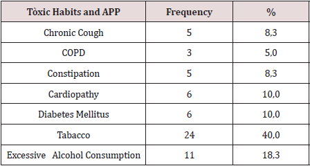
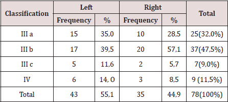
There were operated 78 hernias in 60 patients (18 patients [30.0%] suffered from bilateral hernias, 69 primary hernias and 9 reproduced hernias). The average of age was 55.6 years, the youngest patient was 30 years and the oldest was 77 years, but the majority (12 patients) were in the fifth decade of life. The male sex predominated in 82.9%, which represented a relationship man / woman of 5: 1. 42.9% of patients performed large efforts habitual physicists. The pathological history of the patients (Table 1). It is observed that 24 patients (40.0%) consumed tobacco, and in 11 an excessive consumption of alcohol was collected 18.3%. COPD: Chronic Obstructive Pulmonary Disease (Table 2). The distribution of the series according to the Nyhus classification. Right hernias predominated (55.1%), the indirect variety with large dilation of the ring and destruction of the posterior wall (IIIb) was the most frequent (37 hernias), and 7 femorales hernias and 9 recurrent hernias were operated 73 PET (93.6%) and 5 TAPP (6.4%) were performed. Two of the patients in whom a TEP technique was started were converted to a conventional prosthetic technique by accidental perforation of the peritoneum, passing the CO2 into the peritoneal cavity, and consequently, the loss of the preperitoneal surgical space. Of the 5 TAPP repairs, 3 of them were in the course of a laparoscopic cholecystectomy, and another was the conversion of a failed PET technique. The average surgical time of unilateral hernias was 53.5 min, with a minimum of 25 min and a maximum of 120 min. In bilateral repairs, the average surgical time was 71.3 min, and a minimum of 40 and a maximum was observed. of 110 minutes.

Table 1: Toxicos habits, and personal phalogical history (APP).

lupinepublishers-openaccess-surgery-case-studies-journal

Table 2: Distribution According to the Nyhus Classification.

lupinepublishers-openaccess-surgery-case-studies-journal

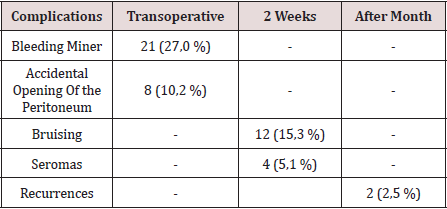
The hospital stay was less than 24 h in 50 patients (71.4%), in 5 it extended from 24 to 48 h, and in 5 to more than 48 hours. The most frequent complication in the transoperative period (Table 3) was minor bleeding in 21 repairs (27.0%) that gave rise to 12 hematomas (15.3%). No complications were observed after the second week, but two patients suffered recurrences (2.5%), more than two months after surgery. The application and evaluation of the VAS scale (Table 4). In the immediate postoperative period, after the patient recovered from anesthesia, 56 individuals (93.3%) were classified as VAS I, and 4 as VAS II. Twenty-four hours after surgery, 14 patients (23.3%) were classified as VAS I, 40 (66.6%) as VASII, 4 patients as VAS III and 2 VAS IV. In the consultation of the first week of postoperatively, 42 patients (70.0%) were classified as VAS I and [10] as VAS II, and two patients with moderate pain (VAS V) appeared in this period. Fifteen days after surgery, 56 individuals (93.3%) were VAS I and a month were 58 (96.6%). The incorporation to the usual activities, including work, was 3 patients a week after surgery, at 15 days they were 19 patients (31.6%) and 54 patients a month (90.0%).

Table 3: Complications.

lupinepublishers-openaccess-surgery-case-studies-journal

Table 4: Evaluation of the Visual Analogy Scale (VAS).

lupinepublishers-openaccess-surgery-case-studies-journal

Discussion

Currently, with the improvement of laparoscopic techniques, these are outlined as safe, reproducible and as a therapeutic option regardless of the age of the patient; nevertheless, the evaluation of the individual must be correct and meticulous in the preoperative period, specifically cardiorespiratory function, since with the TEP method a working space is created between the sheets of the crosssection sheet, richly vascularized, so that absorption and elimination of CO2 is greater than that produced in the peritoneal cavity during the performance of the pneumoperitoneum [10]. Although men predominated, there was a slight increase in women in the series with respect to other authors, [3-5] which could have been due to the inclusion in the study of 3 women who underwent the diagnosis of inguinal hernia, in the course of a laparoscopic cholecystectomy. In laparoscopic practice, it is not uncommon finding of hernia defects diagnosed in the transoperative, in men and women, the latter basically with a history of gynecological disorders. Although the usefulness of hernia repairs in asymptomatic patients is questioned in some articles, [11,12] the authors consider that it would be beneficial for the patient, if conditions permit, the repair of the hernia defect by the TAPP method. The relationship between the hernia disease and the physical efforts, is classic from the Cooper era [1]. In the series, 68% of the patients performed physical activities involving large and medium efforts, and also analyzing the multifactorial character in the pathogenesis of hernia disease, was striking, that approximately half of the patients operated on were smokers, a factor that influences the metabolism of collagen, significantly linked to hernia recurrences [13]. The majority of the repairs were by means of the PET technique, and we consider, as other authors [10,14 -19], that although the TAPP technique brings us closer to the area from a family perspective to the surgeon (peritoneal cavity) and facilitates the so-called “learning curve”, the hernial disease - considering it a parietal defect- should be given solution from this same plane to avoid the likelihood of serious complications of intrabdominal , and to leave the transperitoneal method as a tactical resource when the totally extraperitoneal method is unsuccessful.

The average surgical time was similar to other series [3-6]. It is known that this tends to decrease when the surgical team gains experience [16]. The largest surgical time recorded was in a patient, who was started with a PET technique, but Due to technical difficulties, it was converted to a conventional posterior repair. The fundamental complications were in relation to minor bleeding in the transoperative period and postoperative hematomas. In 3 patients it was necessary to drain the hematoma due to the discomfort caused, however, in the rest of the patients with hematomas and seromas they were treated with conservative measures. In two patients, the recurrence occurred 2 months after surgery, which was interpreted as a technical error. Our results coincide with numerous studies [3-7], that indicate the least postoperative pain of the minimum access techniques, as well as a prompt social and labor reincorporation of the patients. Despite the fact that 70% and 93.3% a week and 15 days postoperatively, respectively, had no pain or discomfort were minimal, only [18] individuals (30%) were incorporated into their usual activities before 15 days. These results contrast with other studies that report a return to work and social activities between 10-15 postoperative days, 4-17- although it is likely that some sociocultural factors are influencing these results. It can be concluded by noting that laparoscopic inguinal hernioplasty is another therapeutic option, mainly in patients with bilateral and reproduced hernias. In the series there were no major transoperative or postoperative complications, only minor bleeding and bruising were present. Most patients were not afflicted by pain 2 weeks postoperatively, however, return to social and labor activities after 15 days was low [18-20].

Read More Lupine Publishers Surgery and Case Studies Journal Articleshttps://surgery-casestudies-lupine-publishers.blogspot.com/


Friday, March 13, 2020

The Optimal Pain Management Methods Post Thoracic Surgery: A Literature Review| Lupine Publishers


Abstract


Post-operative pain control is one of the key factors that can aid in fast and safe recovery after any surgical interventions. Thoracic surgery can cause significant postoperative pain which can lead to delayed recovery, delayed hospital discharge and possibly increased risk of chest complications in the form of atelectasis and even lower respiratory infections. Therefore, appropriate pain management following thoracic surgery is mandatory to prevent development of such morbidities including chronic pain.
Keywords:Thoracic Surgery, Analgesia, VATS, Robotics, Thoracotomy

Introduction

Thoracic surgical procedures can result in severe pain which can present as a challenge to be appropriately managed postoperatively. In particular, thoracotomies are well known for their severity of pain due to the incision, manipulation of muscles and ligaments, retraction of the ribs with compression, stretching of the intercostal nerves, possible rib fractures, pleural irritation, and postoperative tube thoracotomy [1]. Recognition of this has contributed to the development of minimally invasive techniques such as video assisted thoracoscopic surgeries (VATS) and lately robotic surgery [1]. These techniques not only aim to produce better aesthetic results, but also reduce post-operative pain and enhance recovery without compromising the quality of treatment offered. Poor pain management can lead to several and serious complications such as lung atelectasis, hypostatic pneumonia due to avoidance of deep breathing in these patients as a result of pain and superimposed infection [1]. Pain management as a result, does not only lead to greater patient satisfaction, but it also reduces morbidity and mortality in patients undergoing thoracic surgery [2]. Historically, post-operative pain management for thoracic surgery involved the use of narcotics alongside parenteral or oral anti-inflammatory agents [2]. Post chest tube removal patients typically are transitioned to oral analgesia. Multiple additional pain control adjuncts were also implemented with differing levels of success [1]. Over time, intra-operative techniques have been developed which aims to target pain reduction postoperatively [2]. As our understanding of both pain management and the factors that play a role in the development of pain has increased, we have been able to target these and improve postoperative pulmonary morbidity and pain scores [1,2]. We aim to review different means of pain control in this paper in order to assess their effectiveness in achieving optimum results.

Thoracotomy

The mechanism of pain in thoracotomy involves the innervation of the intercostal, sympathetic, vagus and phrenic nerves [3]. Additionally, shoulder pain may result from stretching of the joints during the operation.
After a thoracotomy, pain can persist for two months or more, and in certain incidences it recurs after a period of cessation. The incidence of chronic pain post thoracotomy is reported to be 22-67% in the population [4]. Good surgical technique and effective acute post-operative pain treatment are evident means of preventing post-thoracotomy pain and consequent pulmonary complications [4]. Due to the multifactorial character of the pain, a multimodal approach to target pain is advised. Typically, both regional and systemic anaesthesia are administered. A combination of opioids such as fentanyl or morphine are typically used [5]. A variety of techniques for the administration of local anaesthetics are available at present, and the effectiveness of each is assessed in this paper.
a) Thoracic Epidural Analgesia (TEA)
TEA was the most widely used method of means of analgesia. It was the gold standard means of pain relief [6,7]. It is typically inserted prior to general anaesthesia, at the level of T5-T6, midway along the dermatomal distribution of the thoracotomy incision. A study by Tiippana et al. [8] measured the visual analogue scale (VAS) in order to assess the presence of pain during rest and at the time at which they coughed in 114 patients of whom 89 had TEA and 22 who had other methods of pain control. TEA was effective in alleviating pain at rest and during coughing. In TEA patients, the incidence of chronic pain of at least moderate severity was 11% and 12% at 3 and 6 months, respectively. The study found that at one week after discharge, 92% of all patients needed daily pain medication. The study advised for extended postoperative analgesia for up to the week post-discharge to be administered in order to manage this. The study however concluded overall, that TEA was effective in controlling evoked post-operative pain. However, the study did encounter problems of technical form in 24% of the epidural catheters. The incidence of chronic pain, however, was lower compared with previous studies where TEA was not used. Several other studies support that TEA is superior to less invasive methods. According to Shelley B. et al. [9] TEA was preferred by 62% of the respondents over paravertebral block (PVB) with 30% and other analgesic techniques with 8%. Limitations of this technique included hypotension and urinary retention. Certain patients with active infection and on anticoagulation are excluded from epidural placement.
b) Paravertebral Block (PVB)
PVB is considered an effective method for pain management and its use has been increased in the recent years. This technique involves injecting local anaesthetic into the paravertebral space and it is able to block unilateral multi-segmental spinal and sympathetic nerves. Previous studies have shown that it is effective in achieving analgesia and is associated with a lower incidence of side effects such as nausea, vomiting, hypotension and urinary retention [10,11]. As the lungs are collapsed, it is associated with a lower risk of pneumothorax.
In a study by Davies R.G. et al. [10] there was no significant difference in pain scores, morphine consumption and supplementary use of analgesia between TEA and PVB. The rate of failed technique was lower in PVB (OR =0.28, p=0.007). Respiratory function was improved at both 24 and 48 hours with PVB but only significantly improved at 24 hours.
c) Intercostal Nerve Block (ICNB)
ICNBs are generally administered as single injections at least two dermatomes above and below the thoracotomy incision [12]. It is performed percutaneously or under direct vision, using single injections or through placement of an intercostal catheter. It can also be formed using cryotherapy. It is associated with reduced post-operative pain scores; however, it is less effective than TEA in controlling chronic pain [12]. This was illustrated by a study by Sanjay et al. [12] which found that patients that underwent ICNB had higher pain scores 4 hours post-operatively, than those who received epidural anaesthesia using 0.25% bupivacaine (p<0.05). The study concluded that in the early post-operative period there was significant impact in pain relief for both techniques, but thereafter, epidural anaesthesia was proven to significantly reduce post thoracotomy pain over ICNB. Due to the multifactorial nature of post-thoracotomy pain, various approaches are required in order to target pain. ICNBs are useful in the blockade of intercostal nerves, whilst PVB and TEA appear to block the intercostal and sympathetic nerves. Due to the inability of regional anaesthesia to block the vagus and phrenic nerves which are implicated in the pathophysiology of pain, NSAIDs and opioids are required as adjuncts. TEA is proven to be the most effective means of treating pain alongside PVB; however, it is associated with more side effects than PVB. At present, there are a limited number of studies directly comparing pain control and post-operative outcomes between PVB and TEA. There is no conclusive evidence that either method is superior to the other regarding pain control.

Video-Assisted Thoracoscopic Surgery (VATS)

Existing evidence supports the noninferiority of thoracic PVB when compared to TEA for postoperative analgesia [13]. PVB is versatile and may be applied both unilaterally or bilaterally. It can be used to avoid contralateral sympathectomy, consequently minimising hypotension. This is an apparent advantage it has over thoracic epidural. Furthermore, it offers a more favourable side effect profile when compared to epidural anaesthesia. At present, the factors taken into consideration when selecting a regional technique include tolerance of side effects associated with TEA, consensus on best practice/technique, and operator experience [13]. A randomised controlled trial by KosiÅ„ski et al. [14] compared the analgesic efficacy of continuous thoracic epidural block and percutaneous continuous PVB in 51 patients undergoing VATS lobectomy. The primary outcome measures were postoperative static (at rest) and dynamic (coughing) visual analogue pain scores (VAS), patient-controlled morphine use and side-effect profile. The study found that pain control (VAS) was superior in the PVB group at 24 hours, both at rest (1.7 vs3.3, p=0.01) and on coughing (5.8 vs 6.6, p=0.023), and control of pain at rest was also superior in the PVB group at 36 hours (3.0 vs 3.7 (p=0.025) and at 48 hours (1.2 vs 2.0, p=0.026). There were no significant differences in the postoperative morphine requirements. In regard to side-effect profile, the study showed that the incidence of postoperative urinary retention (defined as no spontaneous micturition for 8 hours or ultrasound-assessed volume of the urinary bladder >500ml) was greater in the epidural group (64.0% vs 34.6%, p=0.0036), as was the incidence of hypotension (32.0% vs 7.7%, p=0.0031). There was no significant difference in the incidence of atelectasis (4.0% vs 7.7%, p=0.0542). However, the incidence of pneumonia was significantly more frequent in the PVB group (3.8% vs 0%, p=0/0331). KosiÅ„ski et al. concluded that PVB is as effective as thoracic epidural block in regard to pain management as it offers a superior safety profile with minimal postoperative complications. A further randomised controlled trial by Okajima et al. [15] compared the requirements for postoperative supplemental analgesia in 90 patients who received wither a PVB or thoracic epidural infusion for VATS lobectomy, segmentectomy or wedge resection. The main outcome measures were pain scores at rest (verbal rating scale 0= none and 10=maximum pain), blood pressure, side effects and overall satisfaction scores relating to pain control (1=dissatisfied and 5=satisfied). The study found a similar frequency of supplemental analgesia (50mg diclofenac sodium suppository or 15mg pentazocine intramuscularly) for moderate pain in both groups, with 56% of those in the PVB group requiring ≥2 doses, compared to 48% in the epidural group (p=0.26). Hypotension, defined as a systolic blood pressure <90mmHg, occurred more frequently in the epidural group (21.2% vs 2.8%, p=0.02). There was no difference in the incidence of pruritus (3.0% vs 0%, p=0.29) and post-operative nausea and vomiting (30.3% vs 25.0%, p=0.62) between both groups. The study found no statistical difference between patient-reported satisfaction in pain control between epidural and PVB using the verbal rating scale (5.0 vs 4.5, p=0.36). The study concluded that PVB offered additional to equivalent analgesia to epidural, a lower incidence of haemodynamic instability postoperatively. A further study by Khoshbin et al. [16] performed an analysis on 81 patients undergoing VATS for pleural aspiration +/- pleurodesis, lung biopsies or bullectomy. The main outcome was postoperative pain levels, documented every 6 hours and scored against the Visual analogue Scale (0= no pain, 10= worst possible pain). In both PVB and epidural groups, bupivacaine 0.125% was the local anaesthetic of choice, with clonidine added to the epidural infusion at 300μg in 500ml. The study showed that there was no significant difference in mean pain scores between PVB or EP (2.1 vs 2.9, p=0.899), therefore concluding that PVB is as effective as epidural in controlling pain post-VATS.

Robotic Lung Surgery

Minimally invasive techniques are considered advantageous over open surgical approaches due to their shorter recovery times, reduced perceived levels of pain post-operatively and shorter postoperative length of stay in hospital [17-19]. Robotic surgery has become a popular method in recent years. Debate remains regarding whether robotic surgery is superior to VATS in regard with pain reduction. A case control study by Louie et al. [19] compared 45 robotic assisted lobectomies (RAL) to 34 VATS lobectomies. The study showed that both groups had a similar mean ICU stay (0.9 vs 0.6 days) and a mean total length of stay (4.0 vs 4.5 days). The study showed that patients that underwent robotic lobectomies had a shorter duration of analgesic use post-operatively (p=0.039) and a shorter time resuming to normal everyday activities (p=0.001). A limitation in this study was an inaccurate record of the amount of pain relief used by the patients, ultimately working as a confounding factor when interpreting the results. In a separate study by Jang et al. [18] 40 patients undergoing RAL were compared retrospectively to 80 VATS patients (40 initial patients and 40 most recent patients), all with resectable non-small cell lung cancer. The study showed that the post-operative median length of stay was significantly shorter in RAL patients compared to the initial VATS patients. The rate of post-operative complications was significantly lower in the RAL group (10%) compared to the initial VATS group (32.5%) and similar to the recent VATS group (17.5%). Post-operative recovery was easier for patients in both the RAL and VATS group due to earlier mobilisation, allowing them to return to their everyday activities quicker. In a retrospective review by Kwon et al. [17] 74 patients undergoing robotic surgery, 227 patients undergoing VATS and 201 patients undergoing anatomical pulmonary resection were assessed and compared with regard to acute (visual pain score) and chronic pain (Pain DETECT questionnaire). The study showed that there was no significant difference in acute or chronic pain between patients undergoing robotic assisted surgery and VATS. Despite no significant difference in pain scores, 69.2% of patients who underwent robotic-assisted surgery felt the approach affected their pain versus 44.2% of the patients who underwent VATS (p=0.0330). These results all support the superiority of robotic surgery over VATS and open approaches with regard to pain, length of hospital stay and recovery times. Both robotic surgery and VATS have their benefits i.e. two-versus three-dimensional view, instrument manoeuvrability, and reduced post-operative pain.

Conclusion

Since post-thoracotomy pain is multifactorial, a multimodal approach is required. In particular, ICNB blocks the intercostal nerves, and PVB and TEA appear to block the intercostal and sympathetic nerves. NSAIDs and opioids are required as valgus and phrenic nerve cannot be blocked by regional anaesthesia. TEA is evident to be the most effective in treating pain alongside with PVB. It is however associated with more side effects than PVB.


Read More Lupine Publishers Journal of Surgery Articles : https://surgery-casestudies-lupine-publishers.blogspot.com/
Read More Lupine Publishers blogger Articles: https://lupinepublishers.blogspot.com


Gallstone Ileus in the Elderly: Still a Challenge, Report of a Case with Review of the Current Literature

  Abstract Introduction:  Gallstone ileus is described as an intestinal obstruction caused by luminal gallstone impaction. It is a mainly ...