Showing posts with label SCSOAJ. Show all posts

Friday, March 24, 2023

Gallstone Ileus in the Elderly: Still a Challenge, Report of a Case with Review of the Current Literature

 Abstract

Introduction: Gallstone ileus is described as an intestinal obstruction caused by luminal gallstone impaction. It is a mainly geriatric disease with a prevalence of over 25 % in the elderly population. Morbidity and mortality rates are high which are caused due to the delayed presentation, diagnosis and treatment in comorbid patients. Interestingly, since the past century, the optimal surgical procedure in this type of mechanical ileus is still highly controversial and challenging.

Presentation of case: In the current paper we demonstrate our experience with a patient treated at our institution due to gallstone ileus. During surgical exploration the impacted stone in the terminal ileum was removed without preforming a cholecystectomy. This decision was made because of the unstable and septic status of the patient necessitating quick relief of the obstruction and no signs of ongoing severe peritonitis and acute Cholecystitis. She had an uneventful postoperative course.

Discussion & conclusion: While enterolithotomy is performed most commonly because of the low incidence of complications, the risk of developing recurrent biliary symptoms has led to a more aggressive approach with concomitant fistula repair and cholecystectomy.

Keywords: Gallstone ileus, elderly patients, surgical therapy, decision-making

Read more about this article click on below link: 

https://lupinepublishers.com/surgery-case-studies-journal/fulltext/gallstone-ileus-in-the-elderly-still-a-challenge-report-of-a-case-with-review-of-the-current-literature.ID.000176.php.

Read more about Lupine Publishers Google Scholar articles 

https://scholar.google.com/citations?view_op=view_citation&hl=en&user=77gTAnQAAAAJ&cstart=20&pagesize=80&citation_for_view=77gTAnQAAAAJ:DUooU5lO8OsC

Monday, November 14, 2022

The Optimal Pain Management Methods Post Thoracic Surgery: A Literature Review


Abstract

Post-operative pain control is one of the key factors that can aid in fast and safe recovery after any surgical interventions. Thoracic surgery can cause significant postoperative pain which can lead to delayed recovery, delayed hospital discharge and possibly increased risk of chest complications in the form of atelectasis and even lower respiratory infections. Therefore, appropriate pain management following thoracic surgery is mandatory to prevent development of such morbidities including chronic pain.

Read more about this article click on below URL: 


Read more about Lupine Publishers Google Scholar Articles: 

https://scholar.google.com/citations?view_op=view_citation&hl=en&user=77gTAnQAAAAJ&cstart=20&pagesize=80&citation_for_view=77gTAnQAAAAJ:PVjk1bu6vJQC

Friday, March 18, 2022

Lupine Publishers | Transfusion Practice in Surgical Resuscitation Department of a University Hospital, Antananarivo (Madagascar)

 Lupine Publishers | Surgery & Case Studies: Open Access Journal

Abstract

Introduction: Given the frequency of blood transfusion in the intensive care unit, it must be performed according to the recommendations to ensure transfusion safety. The aim of our study is to describe the practice of transfusion in a surgical resuscitation service.

Methods: This is a 21-month retrospective and descriptive study (January 2015 - September 2016) in the Department of Surgical Resuscitation of the University Hospital JRA Antananarivo Madagascar. All patients over 15 years old and transfused during the study period are included.

Results: 937 patients were transfused, with a transfusion rate of 25.3%. A male predominance (sex ratio = 2.21) was found with an average age of 45.93 (± 17.74) years. Digestive haemorrhage and postoperative cases are common reasons for transfusion. The blood count and the haemostasis assessment were performed in 58.38% and 50.48% of pre-transfusion patients, respectively. The red blood cell (RBC) is the most used (54.72%), followed by Fresh Frozen Plasma (FFP) (28.68%) and then there have been cases of use of Platelet – Rich Plasma (PRP) (12.93%) and Whole Blood (2.67%). The transfusion event occurred only in 0.43% of patients.

Conclusion: The realities of the country explain the insufficiency of biological arguments for transfusion. And the absence of a well-defined haemovigilance system makes transfusion accidents unrecognized and undeclared. Hence the interest of a transfusion protocol adapted to the Malagasy context.

Introduction

A blood crase disorder is a common occurrence in Intensive Care Unit (ICU) [1]. In general, it is anaemia or a haemostasis disorder. These disorders are at the same time the consequence of the initial pathology motivating the admission in resuscitation but also a factor of aggravation of the pre - existing pathology or the pathology responsible for the admission [2]. To correct these disorders, the use of transfusion is often necessary. But for a good practice of transfusion, like any therapeutic act, it must respect some recommendations [3]. So, this study was done in order to describe the practice of transfusion in intensive care unit of University Hospital /JRA, Antananarivo, Madagascar. accidents, and the blood tests before and after transfusion (haemoglobin, platelet count, prothrombin level). The data were processed by the XLSTAT software version 2019 and the results are expressed as mean with standard deviation, minimum and maximum, and as a percentage.

Results

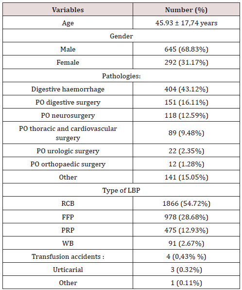
Table 1: General characteristics.
lupinepublishers-openaccess-surgery-case-studies-journal

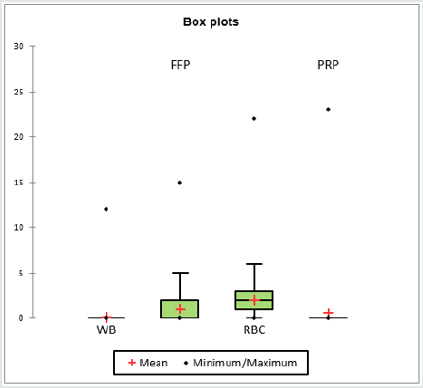
Of the 3700 patients hospitalized in the department during the study period, 937 patients (25.3%) met the inclusion criteria. The average age of these transfused patients was 45.93 (± 17.74) years; with a minimum age of 15, the maximum age was 92. The male gender was predominant (645 patients, 68.83%, sex ratio = 2.21). Digestive haemorrhages and post-operated cases held the top spots in the pathologies presented by these transfused patients, with 43.12% and 41.83% respectively. On the other hand, other pathologies (such as trauma, haemoptysis, haematuria, inflammatory anaemia, leukaemia, various tumours, shock conditions, etc.) were also significant as a reason for blood transfusion with a total of 15.05% (Table 1). Of the 937 patients transfused; before transfusion, 58.38% had a blood count and 50.48% had a haemostasis test; and after the transfusion, only 23.80% had a blood count and the haemostasis assessment was performed in 16% of the patients. The mean haemoglobin level ranged from 92.49 (± 32.49) g/l during the pre-transfusion period to 105.78 (± 22.27) g/l post transfusion. Mean pre-transfusion platelet count was 185.26 (± 117.61) giga / l and 193.70 (± 125.47) giga/l in post transfusion. The prothrombin level (PL) was 62.17 (± 22.74) % before transfusion and 67.13 (± 20.26) % after transfusion (Table 2). Simple transfusion accounted for 99.79% and transfusion exchange 0.21%. The total amount of blood units received by the patients was 3410 with 54.72% red blood cell (RBC), 28.68% fresh frozen plasma (FFP), 12.93% platelet rich plasma (PRP) and 2.67% Whole Blood (WB). Each patient receives on average 2 units of RBC, 1 unit of FFP, 0.5 unit of PRP (Figure 1). There were only 4 cases of transfusion accident including 3 urticarial and 1 dyspnoea andchills.

Figure 1: Variation of transfused LBPs.
Lupinepublishers-openaccess-Surgery-Casestudies

Table 2: Evolution of biological test in pre and post transfusion.

lupinepublishers-openaccess-surgery-case-studies-journal

Hb: Haemoglobin; PLT: Platelet; PL: Prothrombin Level; Pre: Pre-Transfusion; Post: Post Transfusion

Discussion

Resuscitation or intensive care unit is the place where admitted a patient whose vital function is at risk or likely to be. Haematological disorder can enter in this framework of vital function. And often, transfusion is necessary to correct these disturbances. In our case, the frequency of transfusion is 25.3%. In other countries such as Ivory Coast, Denmark, and Canada, they have found transfusion frequency rates of 20%, 25%, and 38% respectively [4-6]. And still according to these studies, these figures are quite high and a policy of reducing consumption of LBP deserves to be implemented. Regarding the profile of transfused patients, there is a male predominance with a low average age (45 years). Unlike those found in studies conducted in occidental countries where the average age is around 60 years [7,8]. The advantage of this relatively low age is the existence of low tolerance thresholds for anaemia; in general, the haemoglobin trigger is 70g/l for a young patient with no antecedent [9]. And for the pathologies presented by these transfused patients, these are the same etiologies that are reported in the literature; that is, trauma, heavy surgery and gastrointestinal bleeding, but of varying proportions [10]. And about the transfusion itself - apart from a simple transfusion, there is, but in a small proportion, the practice of transfusion exchanges in our case. Erythrocyte transfusion exchange is most often indicated during complications of sickle cell disease [11], which is similar to our case. And the use of CGR is the most common in our practice due to the frequency of anaemia during hospitalization in resuscitation. According to the literature, more than 55% of patients admitted to resuscitation have a haemoglobin level of less than 90g/l [10]. In our case, the average pre-transfusion haemoglobin level was 92.49 g / and there was a one-point post-transfusion gain for the haemoglobin level. It should be noted that the pre- and post-transfusion biological assessment rate in our practice is quite low. Since the biological assessment for deciding a transfusion or not is not always available, a study, similar to that carried out by Kranenburg FJ et al. [12] who sought predictive factors for transfusion, deserves to be instituted. This is in order to properly codify the transfusion practice by limiting the consumption of LBP and avoiding its complications. However, these complications can be dangerous and engage the prognosis of the patient [13]. In our case, the frequency rate of these complications is abnormally low. Three contingencies can explain this Figure 1, so that the frequency of these accidents is so low, it would require a quality technical platform and a good haemovigilance system; which is not our case. The other eventualities would be that other cases of transfusion complications exist but remain undeclared or unknown, hence the interest of protocolisation of transfusion safety [14].

Conclusion

This study allowed us to profile transfused patients and describe the practice of transfusion in a surgical resuscitation of a university hospital in Madagascar. In this time of LBP shortage [15], this study could improve transfusion practice not only by streamlining the use of PSLs, but also to limit transfusion accidents.

Read More Lupine Publishers Surgery and Case Reports Journal Articles: https://surgery-casestudies-lupine-publishers.blogspot.com/

Thursday, February 20, 2020

Acute Liver Failure and Thyrotoxicosis Managed with Liver Transplant and Thyroidectomy- Lupine Publishers

Abstract

Acute liver failure and hyperthyroidism are not typically common, although some cases have been reported. The mechanisms involved and optimal management are not well-defined. This article presents the case of a 32-year-old African American female referred for evaluation of abdominal pain and jaundice, with a past medical history of systemic lupus erythematosus and Grave’s disease. She had thyrotoxicosis after administration of contrast and developed acute liver failure culminating in liver transplant and subsequent total thyroidectomy with a favorable outcome.
Keywords: Grave’s Disease; Autoimmune Hepatitis; Thyrotoxicosis; Acute Liver Failure; Liver Transplant; Thyroidectomy; Contrast Induced
Abbrevations: OLT: Orthotopic liver transplant; SLE: Systemic Lupus Erythematosus; TT: Total thyroidectomy; HD: Hospital day; POD: Post-operative day; ANA: Anti-nuclear antibodies; NaMELD: Sodium model for end-stage liver disease; PTU: Propylthiouracil.

Introduction

The association between liver function abnormalities and thyroid dysfunction is well established dating back to at least 1874 when Habershon presented a case of exophthalmic goiter and jaundice to the London Medical Society [1]. The range of liver dysfunction can go from mild elevation of hepatic enzymes to acute liver failure, which is defined as the presence of hepatic encephalopathy within 8 weeks of initial symptoms in a patient without underlying liver disease. Multiple scales have been devised to predict mortality and determine which patients would benefit from orthotopic liver transplant (OLT), with the presence of hepatic encephalopathy as a key indicator [2]. Here we present a case of a 32-year-old female with systemic lupus erythematosus (SLE) and exacerbation of Grave’s disease after iodinated contrast that presented with acute liver failure and thyrotoxicosis requiring OLT and total thyroidectomy (TT) after medical management of both conditions.

Case

A 32-year-old African American female was referred to the emergency room for evaluation of a 3-week history of worsening abdominal pain and jaundice. She also reported loss of appetite and occasional epigastric discomfort. Her past medical history included SLE and Grave’s disease [3]. Both conditions were well controlled, and her medications included multivitamins, hydroxychloroquine and ferrous sulfate. Three weeks prior to admission she underwent a CT scan with iodinated contrast for abdominal pain at an outside hospital. At that time, she received prednisone (60mg PO qd) and diphenhydramine for a presumed diagnosis of autoimmune hepatitis with positive antinuclear antibodies (ANA) at 1:320. Ferritin, alpha 1 antitrypsin, acetaminophen and viral serologies were all negative prior to admission. Her initial vital signs showed BP 110/70 mmHg, HR 134 bpm, RR 20 bpm, T 37°C and SpO2 100%. Physical examination was significant for generalized jaundice and moderate abdominal pain on palpation.
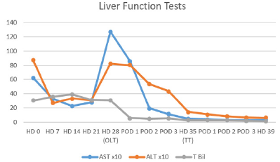
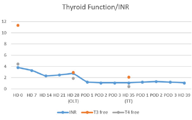
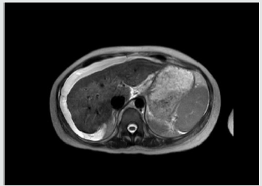
At that time her laboratories were INR 3.8, WBC 36.6 k/uL, K 2.7 mmol/L, lactate 2.4 mmol/L, Alk Phos 187 IU/L, AST 625 IU/L, 872 IU/L, Built 30.2 mg/dL, Bild 21.9 mg/dL, T3 11.3 pg/mL, T4 4.46 ng/dL, TSH 0.02 uIU/mL and ceruloplasmin 26 mg/dL. On HD #11 her 24hr copper excretion was 241.6 ug/d. Her admission Named was 35 and 31 at time of transplant. Renal function remained normal throughout her hospitalization. Trends for her hospital stay are shown in (Tables 1 & 2) After initial stabilization, Doppler ultrasound revealed patent vasculature, and MRI showed nodular appearance of the liver consistent with edema or early cirrhosis (Figure 1). The liver transplant service was consulted and a standard workup for potential liver transplant was started.
Table 1: Liver Function Tests. OLT: Orthotopic Liver Transplant. TT: Total Thyroidectomy.
Lupinepublishers-openaccess-Surgery-Casestudies
Table 2: TSH remained undetectable at <0.02.
Lupinepublishers-openaccess-Surgery-Casestudies
Figure 1: MRI of abdomen showing nodular appearance of liver consistent with edema or early cirrhosis.
Lupinepublishers-openaccess-Surgery-Casestudies
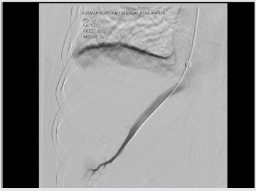
A transvenous liver biopsy on HD #6 was positive for marked active hepatitis with bridging necrosis, parenchymal collapse and minimal focal staining for copper. At the time of biopsy, a hemodynamic study was performed consistent with portal hypertension (Figure 2). Blood cultures were positive for E. coli and urine cultures for E. faecalis on admission, and she received ceftriaxone and meropenem with negative conversion of cultures. Her condition continued to deteriorate with worsening hepatic encephalopathy despite optimal treatment with rifaximin and lactulose. Oral prednisone was continued at 40 mg PO/qd. Her thyrotoxicosis was managed with propranolol and potassium iodide oral solution. An echocardiogram ruled out congestive heart failure. At HD #26 she was admitted to the intensive care unit and due to her condition, it was deemed she was at risk of death within one week and was listed as emergency status 1A on the united network for organ sharing.
Figure 2: Transvenous hemodynamic study consistent with portal hypertension. Free hepatic pressure 19 mmHg, wedge hepatic pressure 26 mmHg, hepatic venous gradient 7 mmHg.
Lupinepublishers-openaccess-Surgery-Casestudies
A suitable donor became available on HD #28 and she underwent OLT with methylprednisolone and mycophenolate induction without complications. She received 250 mcg of potassium iodide the night prior to transplant. Liver explant revealed extensive bridging necrosis, parenchymal collapse, no definite areas of fibrosis and minimal copper staining. She recovered without incidents from transplant and underwent total thyroidectomy on POD #7, she was then started on levothyroxine. She was discharged on HD #39 to an acute rehabilitation facility. At 1-year follow up, she has excellent graft function, remains euthyroid and has not experienced flare ups of SLE.

Discussion

Exposure to iodinated contrast is well known to cause thyroid dysfunction, with an iodine content of 320 to 370 mg/mL it is well above the recommended daily allowance and can precipitate thyrotoxicosis via the Jöb-Basedow phenomenon in patients with Grave’s disease [3]. Hyperthyroidism, particularly in Grave’s disease, is associated with primary biliary cirrhosis and autoimmune hepatitis. In patients without heart failure and hyperthyroidism, the liver biopsy has demonstrated fatty infiltration, cytoplasmic vacuolization, nuclear irregularity and hyperchromatism [4]. Although the exact mechanism for the observed liver-thyroid interactions is unknown, the following have been proposed:
a. systemic effects of thyroid excess,
b. direct toxic effects of thyroid hormone,
c. intrinsic liver and thyroid autoimmune mechanisms,
d. abnormal thyroid metabolism due to liver disease, and
e. subclinical physiologic effects of thyroid hormone [5].
feasible explanation is the presence of a hypermetabolic state with increased hepatic oxygen consumption, but without increases in hepatic blood flow, affecting the centrilobular zones and interfering with bile transport. These findings are consistent with the picture of cholestasis usually present in such cases [6]. In a series of 84 patients with acute liver failure, Anastasiou and colleagues reported a 50% incidence of thyroid hormone abnormalities, with a worse outcome in patients with hyperthyroidism, believed to be secondary to an increase in oxygen consumption and decreased organ perfusion [7]. In the current case, the presence of drug-induced liver injury had to be ruled out, hydroxychloroquine has been associated with acute liver failure requiring liver transplantation, but it appears within two weeks of starting therapy [8]. There is a well-known correlation between anti-thyroid medications and hepatic dysfunction, for example, propylthiouracil (PTU) and methimazole are associated with an incidence of severe liver injury in 0.1% and 0.1-0.2% of patients, respectively [9].
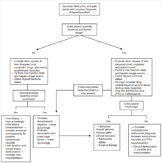
Figure 3: Proposed algorithm for management of patients with symptoms of liver dysfunction and previous diagnosis of hyperthyroidism. [Figure note: Figure obtained from de Campos Mazo, 2013; © 2013 Mazo et al, publisher and licensee Dove Medical Press Ltd. This is an Open Access article which permits unrestricted noncommercial use, provided the original work is properly cited.] [10].
Lupinepublishers-openaccess-Surgery-Casestudies
To our knowledge, our patient was not exposed to these medications. In the absence of established guidelines, a useful algorithm for the management of liver dysfunction with hyperthyroidism has been proposed, (Figure 3) This algorithm is based on a case series of 8 patients in which two cases developed acute liver failure suitable for OLT [10]. Nonetheless, the timing and sequence of OLT and thyroidectomy is not well established. Although it has been proposed not to delay the thyroidectomy as it might represent a potentially life-saving procedure [11]. The incidence of acute liver failure in the setting of hyperthyroidism remains a rare event, with patients undergoing OLT and thyroidectomy even more unusual. In this case report, adequate control of the thyrotoxicosis was achieved prior to transplant with oral potassium iodide and propranolol. In this patient with rapidly progressive liver failure, it appeared clinically prudent to undergo emergent OLT followed by TT.

Conclusion

The presence of ANA in a patient with SLE and Grave’s disease suggest the diagnosis of autoimmune hepatitis, however in this case report, the administration of iodinated contrast and the presence of abnormal copper metabolism made the diagnosis more difficult. The optimal management of these patients is not well established, with only a few cases reported in the literature. Based on our experience, medical stabilization followed by OLT and TT appears to have been an adequate course of treatment. Further studies are needed to help determine guidelines./p>


To know more about Open access publishers click on Lupine Publishers


Gallstone Ileus in the Elderly: Still a Challenge, Report of a Case with Review of the Current Literature

  Abstract Introduction:  Gallstone ileus is described as an intestinal obstruction caused by luminal gallstone impaction. It is a mainly ...