Lupine Publishers | Surgery & Case Studies: Open Access Journal
Acquired digital fibrokeratomas are a benign soft tissue tumor with
typical appearance and anatomical locations. This lesion
generally occurs in middle aged males with common sites of occurrence in
the digits of upper or lower extremities. Previous case
studies have reported incidences of this lesion appearing on heels of
middle-aged males but are generally described as giant digital
fibrokeratoma based on the lesion’s diameter. This case report describes
an acquired digital fibrokeratoma in a pediatric female
patient in an infrequent location.
Keywords: Acquired digital fibrokeratoma; Pediatric; Heel
Introduction
Acquired digital fibrokeratoma is a rare benign soft tissue
tumor typically presenting on the hands and feet of middle-aged
males. Clinical examination tends to reveal a solitary, round, firm,
skin colored lesion less the 1 centimeter in diameter with either
a sessile, dome-shaped or pedunculated base [1-4]. The patient’s
history could entail a slowing-growing lesion without any known
traumatic event which becomes painful upon compression through
enclosed shoes. Diagnosis of the lesion is accomplished through
clinical and physical examination, various biopsy techniques
and histological and surgical pathology evaluation. Treatment
is dependent on the patient’s desired outcomes. Conservative
treatment entails offloading the lesion through various pads,
proper shoe wear, or topical anesthetics. Surgical intervention
entails removal of the lesion en-toto. Surgical intervention has been
favored in recent times as the reoccurrence rate after excision has
shown to be rare [1,2,5]. This case report describes an acquired
digital fibrokeratoma in a pediatric female patient in an infrequent
location.
Case Report
A 13-year-old female with past medical history significant for
asthma, allergic rhinitis, atopic dermatitis, and eczema presented
to clinic for a painful right heel lesion. The painful heel lesion began
several months prior without any known traumatic events. Per the
patient, the lesion began as a callus but progressed in size over the
following months. Pain occurred with direct pressure secondary
to enclosed shoe wear. Clinical examination revealed a firm, nonmobile,
7mm circular skin lesion with a pedunculated based
located on the posterior aspect of the heel. Previous treatments
of offloading pads and topical callus remover were ineffective.
Surgical intervention was warranted due to failed conservative
treatments and an MRI with and without contrast was obtained
to further evaluate the lesion. Obtained MRI showed a soft tissue
mass involving the dermis and epidermis along the posterior heel
without extension into underlying osseous, ligaments or tendon
structures. An excisional biopsy was planned for removal of the
skin mass. Under monitored anesthesia care a 3:1 elipse incision
was made which encompassed the skin lesion in total. The incision
was deepened into the subcutaneous layer and was excised in a
full thickness flap. The proximal pole of the lesion was tagged with
a 4-0 prolene suture and sent for pathological and histological
examination. The wound was closed in usual manner, with sterile
dressing application. She was allowed to weight bear in a CAM boot
and seen in office on post-operative day 10. At post-operative visit
one, she reported no pain, the surgical incision was well-coapted
and sutures were removed. She was instructed to transition out of
the CAM boot and into regular shoes over the following week and
given a 4 week follow up appointment. She canceled her second
post-operative appointment and was not seen in clinic again.
Pathological report obtained confirmed a diagnosis of acquired
digital fibrokeratoma.
Discussion
In 1965, Steel published case reports on an unspecified
periungual fibrous tumor which he described as a garlic-clove
fibroma. Bart et al. reported on ten acral tumors in 1968 which were
deemed similar to Steels previous findings but also resembled a
“rudimentary supernumerary digit” with distinct histopathological
findings and sites of occurrence. They termed the lesion as an
acquired digital fibrokeratoma though suggested the lesion was
not a fibroma rather a protrusion of the dermis [6,7]. Verallo et al
also reported on 32 cases of similar lesions in 1968 as described
by Bart et al. but with the expectation of six additional sites of
occurrence. They suggested to omit the term “digital” from the
name as described by Bart et al. to merit descriptive clarity. Reed et
al. suggested the term acral to be utilized in describing the location
of acquired fibrokeratomas. Recent case reports have described
various locations of this lesion which lead authors to evolve the
lesions descriptive terminology to acral fibrokeratoma [8-16].
Yi-Chiun Tsai et al. retrospectively reviewed 124 patients with
a histopathological diagnosis of acquired digital fibrokeratoma
over a 13-year period to characterize the distribution and surgical
outcomes of these lesions. They found the mean age of occurrence
to be 42 years, with a male predilection of 2:1, and 30/124 (24%)
of the lesions located on nondigital areas. Overall recurrence rate
after surgical resection was 5/124 (4%). Due to a low recurrence
rate, surgical resection is generally recommended in treatment of
these lesions. One most obtain a thorough history, clinical, physical,
and histological evaluation for accurate diagnosis. Patients history
general describes a slow growing lesion, without known trauma,
which may become painful with compression. Physical examination
reveals a small, firm, solitary, painless, skin tone color lesion which
can arise in various locations. Under dermoscopy observation
one visualizes a homogenous pale-yellow center surrounded by
a hyperkeratotic scaly collarette with globular vessels located in
the periphery of the lesion [16]. Histologically evaluation shows
a benign fibroepithelial tumor, with acanthotic epidermis and
thickened, often, branching rete ridges. The lesions core is formed
by closely packed and interwoven collagen bundles which are
generally vertically oriented. Elastic fibers are sparse but often
the lesion is highly vascularized [1-4,17-19]. Differential diagnosis
includes supernumerary digits in pediatric patients, cutaneous
horns, pyogenic granuloma, exostosis keloid, dermatofibrosarcoma,
eccrine poroma, neurofibroma and verruca [1,5,18]. The
pathogenesis of acquired digital fibrokeratoma is unknown though
mechanisms of occurrence have been proposed. Injury and minor
trauma have been hypothesized as triggering factors, but case
reports have not supported these mechanisms [1,18,20,21]. Kint et
al. suggested neoformation of collagen produced acquired digital
fibrokeratoma lesions based on histological findings of denser
collagen fibers with capillaries and fibroblasts. They also described
three types based on clinical and histological findings. Type I is
noted to have a dome-shape and contains fibroblast between
collagen bundles with fine elastic fibers and numerous capillaries
in the dermis. Type II is a taller and hyperkeratotic lesion which
contains more fibroblast and less elastic fibers then type I. Type
III can be flat or dome-shaped and is defined by poor cellular
structures and no elastic fibers [18].
In this case, the patients age, sex, and lesion location were
atypical. Previous published case studies have reported on acquired
digital fibrokeratoma located on a patient ‘s heel but are generally
observed in middle aged males and described as giant acquired
digital fibrokeratomas [10,12,13,20,22]. Majority of acquired
digital fibrokeratoma measure less than 1cm in diameter. When
lesions are greater than 1cm, they are described in the literature
as a giant acquired digital fibrokeratomas [10,12,23,24]. To our
knowledge, this is the first case report describing an acquired
digital fibrokeratoma in a female pediatric patient located on the
heel.
Read More Lupine
Publishers Surgery and Case Reports Journal Articles: https://surgery-casestudies-lupine-publishers.blogspot.com/



 is the relative displacement of the cross section of the
electro elastic actuator along
is the relative displacement of the cross section of the
electro elastic actuator along  axis,
axis,  is the control parameter in
the form the electric field strength of the electro elastic actuator
along i axis on the interval [0,t], the initial value of the electric
field strength
is the control parameter in
the form the electric field strength of the electro elastic actuator
along i axis on the interval [0,t], the initial value of the electric
field strength  the value of t, the initial value the relative
displacement
the value of t, the initial value the relative
displacement  and the sign of the rate
  of the electric field
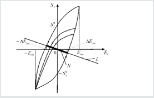
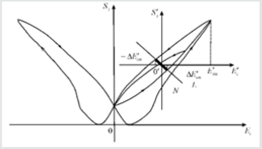
strength variation. Let us consider hysteresis characteristic of the
deformation of the electro elastic actuator on (Figure 1). The set
of the electric field
strength variation. Let us consider hysteresis characteristic of the
deformation of the electro elastic actuator on (Figure 1). The set
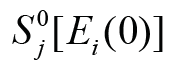 the vertical segment
 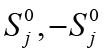 bounded by the points of
intersection of the ordinate axis with the hysteresis loop at the
maximum admissible field strength in the piezoactuator. We receive
for the stable linear part of the control system the stationary set N
and the equation for the straight-line L has the form
bounded by the points of
intersection of the ordinate axis with the hysteresis loop at the
maximum admissible field strength in the piezoactuator. We receive
for the stable linear part of the control system the stationary set N
and the equation for the straight-line L has the form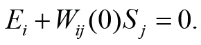
 of the linear part of the control system. The set of points N for
intersection of this straight line L with the hysteresis characteristic
represents the segment of the straight line marked. At
of the linear part of the control system. The set of points N for
intersection of this straight line L with the hysteresis characteristic
represents the segment of the straight line marked. At 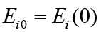 and
and
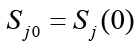 we have the stationary solution to the control system with
hysteresis on Figure 1. We obtain the stationary set in the form of the
marked segment N of straight-line L in Figure 1 with the set
of pairs
we have the stationary solution to the control system with
hysteresis on Figure 1. We obtain the stationary set in the form of the
marked segment N of straight-line L in Figure 1 with the set
of pairs 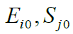 The set of points N for intersection of this straight
line L with the hysteresis characteristic. For the stationary set the
equation has the following form
The set of points N for intersection of this straight
line L with the hysteresis characteristic. For the stationary set the
equation has the following form 
 the hysteresis loop of the piezoactuator and the quantities of
the derivative
the hysteresis loop of the piezoactuator and the quantities of
the derivative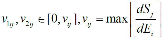

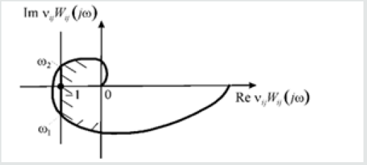
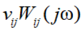
 is the amplitude-phase characteristic of the
open-loop system and in brackets j is the imaginary unity and
is the amplitude-phase characteristic of the
open-loop system and in brackets j is the imaginary unity and 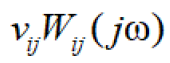 is the frequency The amplitude-phase characteristic of the openloop
system
is the frequency The amplitude-phase characteristic of the openloop
system  on (Figure 3) should be situated to the right
of the straight line
on (Figure 3) should be situated to the right
of the straight line