Showing posts with label Journal of Case Studies. Show all posts

Friday, October 25, 2019

Lupine Pubishers| Cervical Spinal Cord Injury During Coronary Artery Bypass Graft Surgery in An Elderly: A Case Report

Lupine Publishers | Journal of Surgery & Case Studies


Case Report


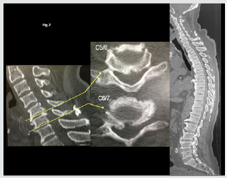
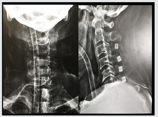
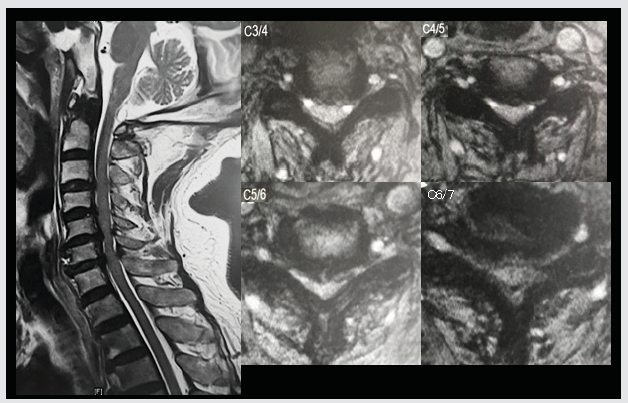
Intraoperative spinal cord injury (SCI) is uncommon and the pathology has not cleared. We report cervical cord injury without vertebral fracture during a coronary artery bypass operation. Cervical cord compression (CCC) is compression of the cervical spinal cord that occurs during the normal course of aging and may progress into cervical spondylotic myelopathy (CSM), which can cause neurologic dysfunction. Many people with cervical cord compression are asymptomatic. However, patients with CCC are at higher risk of SCI following minor injury. An 85-year-old woman with asymptomatic cervical cord compression underwent a coronary artery bypass graft surgery. After the surgery, he developed tetraplegia. MRI revealed severe spinal cord compression from C3 to C7 and C5/6 signal change of spinal cord (Figure 1). The radiograph and CT showed diffuse idiopathic skeletal hyperostosis and thoracic kyphosis of 55 degree (Figure 2). Cervical SCI after was diagnosed. After the diagnosis, we underwent laminoplasty (Figure 3). And he almost completely recovered after surgery.
Figure 1: Magnetic resonance imaging (MRI) of the cervical spine.
Lupinepublishers-openaccess-Surgery-Casestudies
Figure 2: MRI showed multilevel spinal cord compression.
Lupinepublishers-openaccess-Surgery-Casestudies
Figure 3: The radiograph and CT showed diffuse idiopathic skeletal hyperostosis and thoracic kyphosis Radiograph after laminoplasty for cervical spinal cord injury.
Lupinepublishers-openaccess-Surgery-Casestudies
Iatrogenic cervical SCI after non spinal surgeries that requires neck hyperextension is rarely reported, probably due to underdiagnosis and underreport. In the current aging society, the prevalence of patients with CCC is expected to increase. In our population-based magnetic resonance imaging (MRI) study, the prevalence rate of CCC was 24.4%, and it was higher with increasing age in both men and women [1]. Among the 15 cases (including ours) published in the literature [2], most patients had cervical cord compression and were old men. Eight patients had undergone coronary artery bypass surgery that requires prolonged operative time. Only 3 patients had almost complete recovery. Two patients required tracheostomy for long-term ventilator support. Two patients died. These cases reiterate the potential risk of iatrogenic SCI in people with predisposing conditions such as cervical spondylosis and CCC. Moreover, in this case, the patient had diffuse idiopathic skeletal hyperostosis and thoracic kyphosis of 55 degree, which can lead inflexibility of spine. The inflexibility of thoracic spine might cause hyperextension of cervical spine during the surgery. Surgeries requiring prolonged neck hyperextension put patients with cervical spondylosis and CCC at risk for SCI. It is essential to recognize the potential occurrence of iatrogenic SCI might endanger patients’ lives.

For more Lupine Publishers Open Access Journals Please visit our website:
For more Surgery Journal articles Please Click Here:
To Know More About Open Access Publishers Please Click on Lupine Publishers

Follow on Linkedin : https://www.linkedin.com/company/lupinepublishers
Follow on Twitter   :  https://twitter.com/lupine_online

Wednesday, August 28, 2019

Lupine Publishers | Surgical Stabilization of Rib Fractures: Emerging Indications

Lupine Publishers | Journal of Surgery & Case Studies


Abstract


Introduction: Rib fractures are a common injury after road traffic accidents. While most simple rib fractures heal well, multiple rib fractures may result in acute life-threatening complications or chronic disability and work loss. Though surgical fixation of rib fractures has most commonly been restricted to multiple rib fractures with flail chest, there has been a recent interest in fixation of multiple rib fractures with chest deformity to preclude chronic disability and loss of work.
Case Report: We report the case of a 34 year male with multiple rib fracture and chest deformity due to multiple, displaced fractures of 3rd to 10th ribs on the left side. He was treated with open reduction and internation fixation of ribs with 2.4mm titanium reconstruction plates and screws. The emerging indications of rib fracture fixation, as seen in this patient, are discussed.
Conclusion: Longer duration of hospital stays and delay in returning to normal life result in poor quality of life and add to direct and indirect treatment expenses. A case-based approach is essential in the decision-making for surgical fixation of multiple displaced rib fractures.
Keywords: Rib Fractures; Fracture Fixation; Chest Deformity


Introduction

Rib fractures are one of the most common injuries after road traffic accidents. Most simple rib fractures heal well with minimum intervention. But multiple rib fractures may require use of mechanical ventilation and sometimes surgical management [1]. Thoracic trauma comprises 10-15 % of all trauma and are the causes of death in 25 % of all fatalities due to trauma [2]. We present a case of multiple rib fractures and chest deformity and present the outcome of surgical fixation and its significance.


Case Summary

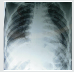
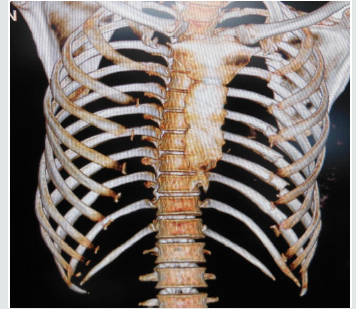
A 34 year male, a bus conductor, was brought to our hospital in the emergency room with an alleged history of road traffic accident. He sustained mild head injury with a history of loss of consciousness and there were multiple abrasions all over his body. He complained of severe excruciating pain during breathing and movements of left arm, with a pain score in VAS scale at 8-9(0-10). Pain was nonresponsive to analgesics. He had significant depression of the chest wall on the left side; chest wall movements were equal bilaterally. Computed tomography of the brain showed no parenchymal injury. Plain chest radiograph (Figure 1) and computed tomography with 3D reconstruction (Figure 2) demonstrated multiple, displaced fractures of 3rd to 10th ribs on the left side. There was no evidence of pneumothorax, hemothorax or lung contusional injury.
Figure 1: Anteroposterior radiograph of the chest demonstrating fracture of 3rd to 11th ribs.
Lupinepublishers-openaccess-Surgery-Casestudies
Figure 2: 3-D computed tomography demonstrating the displaced fractures.
Lupinepublishers-openaccess-Surgery-Casestudies
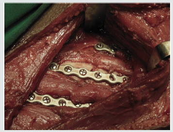
Figure 3: Intraoperative picture demonstrating placement of 2.4 mm titanium reconstruction plates and screws to fix the fractures.
Lupinepublishers-openaccess-Surgery-Casestudies
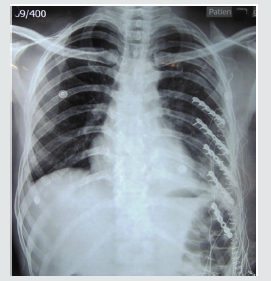
Figure 4: Postoperative radiograph showing surgical fixation of 6th to 10th ribs.
Lupinepublishers-openaccess-Surgery-Casestudies
Considering the presence of chest wall deformity and multiple consecutive rib fractures, surgical stabilization of the ribs was planned. Under general anesthesia, patient was positioned on left lateral position, and through a single lazy- S incision starting from lower border of scapula with a length of 6 cm, lattisimus dorsi muscle was exposed and split along the fibers and access to the ribs was made by stripping off the intercostal muscles. The 6th to 10th ribs were reduced and fixed with 2.4 mm titanium reconstruction plates and screws (Figures 3 & 4).
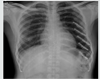
There were no signs of pleural tear after fixation, as clinically confirmed by positive pressure ventilation. The wound was closed in layers with a vacuum drain in-situ. He made a rapid recovery with marked reduction in his pain and discomfort (VAS score of 5) on post-operative day 1. The chest wall deformity was fully corrected. He was discharged on the 3rd post-operative day. Patient was last followed-up at 7months. The fractures had united (Figure 5) and recovery was uneventful. He had returned to work 3weeks following surgery.
Figure 5: Anteroposterior chest radiograph at 7 months following surgery showing fracture consolidation.
Lupinepublishers-openaccess-Surgery-Casestudies


Discussion

Incidence of rib fracture reported by various studies ranges between 7 - 40 %. Most commonly 4th - 9th ribs are fractured. Fractures of upper ribs (1st & 2nd) usually signify severe trauma with increased risk of great vessel injuries [2]. Recently there has been a resurgence of interest in the surgical management of rib fractures [3,4]. Indications for surgical fixation of rib fractures include flail chest, severe chest wall deformity, failure to wean from mechanical ventilation, chronic pain or disability, pulmonary herniation, nonunion and “on the way out” after thoracotomy [5]. Initial research suggests that in select patients, operative management of chest wall injuries is a promising treatment option. Granetzy et al. [4] in 2005 randomised 40 patients who experienced fractures of 3 or more ribs to receive either conservative or surgical treatment and the results showed that patients in the surgical group experienced significantly fewer days on mechanical ventilation, decreased stay in the Intensive Care Unit and hospital stay and less restrictive pattern on pulmonary function tests 2 months after treatment [6]. Similar results were found by Nirula et al. [5] in 2006 where they treated 60 patients with rib fractures [7]. Favourable long term outcomes of patients undergoing surgical chest wall stabilization was documented from a prospective study by Lardinois et al. [8], who had done surgical stabilization of 60 patients of chest wall injuries from 1990-1999.
Rib fractures have been associated with significant disability and loss of work [9]. Hence selected patients with multiple rib fractures but without flail chest have been hypothesized to benefit better from open reduction with internal fixation than from nonoperative treatment [10,11]. All existing surgical indications are relative. Surgical repair has been attributed to possible sooner return to work and usual activities [5,12]. In a retrospective study by Solberg et al on 16 patients of unilateral rib fracture and chest wall deformity, the overall recovery of the surgically treated patient was much earlier than that of those who were treated conservatively [13]. However, no cohort study is available to confirm the beneficial effects of surgical fixation for multiple rib fractures without flail chest [5,12]. Treatment must be individualized on the basis of the patient’s fracture pattern, overall medical condition, and functional status [12]. This patient presents an ideal scenario where a surgical fixation of the rib fracture would result in better clinical outcomes and reduce the morbidity of prolonged pain and disability and loss of work.


Conclusion

The most preferred modality of treatment of rib fractures is non-operative, with analgesics and active chest physiotherapy. However recovery is prolonged or associated with complications, especially in the presence of multiple rib fracture, floating ribs or a flail chest. Longer duration of hospital stay and delay in returning to normal life also result in poor quality of life and add to direct and indirect treatment expenses. Hence, it is rational to manage certain patients with multiple rib fracture surgically to reduce morbidy, mortality and loss of work. Clinical message: The report stresses the need to make a case-based approach in decision-making and the need to have a lower threshold for surgical fixation in the presence of multiple displaced rib fractures. Further cohort studies are needed to confirm the benefits of internal fixation of multiple rib fractures in the absence of flail chest.
For more Lupine Publishers Open Access Journals Please visit our website:
For more Surgery Journal articles Please Click Here:
To Know More About Open Access Publishers Please Click on Lupine Publishers

Friday, June 21, 2019

Lupine Publishers - Journal of Surgery

Dental Prosthetic Rehabilitation of a Tumor Patient AfterRadiotherapy and Mandibular Resection by Soo Jeong Portscher Kim in Surgery & Case Studies Open Access Journal in Lupinepublishers

Background: Many patients with a history of head-and-neck tumors present with hard- and soft-tissue defects resulting from their disease and treatment that will preclude any dental prosthetic rehabilitation without the use of implants.
Case Report: A 57-year-old male patient with a history of tonsil and piriform sinus carcinoma sought prosthetic rehabilitation after successful radio-/chemotherapy and local mandibular resection. His dentition was severely reduced, consisting of inadequate crown restorations in the maxillary anterior segment and the mandibular right canine. The treatment decision was to rehabilitate the maxilla conventionally and the locally resected mandible with an implant-supported telescopic denture after extraction of tooth 43. Four implants were placed (32, 33, 42, 43) and allowed to osseointegrate submerged for 3 months, followed by surgical exposure, tightening abutments, and cementing conical crowns that served as primary telescopes. The secondary telescopes consisted of electroplated copings which were intraorally bonded to the tertiary structure, thus ensuring a perfect fit of the denture.
Conclusion: Other options for denture retention were contraindicated: ball or Locator attachments because of the existing xerostomia and pain from the resection-related scar tissue, and a bar due to its higher demands on self-performed oral hygiene and very high requirements for extending the superstructure in the event of an implant loss. The telescopic denture we provided has none of these drawbacks, nor does it require any extensive cantilever saddles because the masticatory loads are largely absorbed by the implants with almost no force transmission to the mucosa. The only treatment-related complication was a bony dehiscence, which was successfully managed by slightly reducing the bone and harvesting two connective-tissue grafts from the palate to close the perforation. After one year, the denture continues to be intact and functional. The patient reported a short period of adaptation followed by excellently restored function and greatly improved phonetics.


For more Lupine Publishers Open Access Journals Please visit our website:
For more Surgery Journal articles Please Click Here: 


Wednesday, June 19, 2019

Lupine Publishers - Journal of Surgery

Intellectual Corruption at the APA by James F. Welles in Surgery & Case Studies: Open Access Journal in Lupinepublishers

A book I wrote, Understanding Stupidity, was reviewed by Dr. Thomas O. Blank of the School of Family Studies and Center on Aging at the U. of Connecticut, Storrs. The review appeared in the Sept. 1993 issue of Contemporary Psychology, a journal of reviews published by the American Psychological Association (APA). It was very harsh, in some ways un professional and demonstrated Dr. Blank’s inability to understand what he had read. I availed myself of the opportunity to respond in the “Point/ Counterpoint” format made available to aggrieved authors by the journal. This consisted of a four-part exchange of statements between myself and Dr. Blank and appeared in the May 1994 issue. In his last comment, to which I had no opportunity to reply in print, he alleged that my stated view “Normal human behavior is not necessarily adaptive” could be accessed (i.e., found) elsewhere. I received an advanced copy of this statement and twice challenged him in writing to document his claim but received not even the courtesy of a reply. In April, I wrote Dr. John Harvey, the editor of the journal, asking him to see to it that Dr. Blank document his claim or that an appropriate correction be published in a forth coming issue. He was explicit in his refusal to do anything to resolve the matter. I then turned to the APA and had a number of phone con variations and exchanged letters with Leslie Cameron, Director, APA Journals, in May and June regarding this matter, all to no effect.


For more Lupine Publishers Open Access Journals Please visit our website:
For more Surgery Journal articles Please Click Here: 

Monday, March 25, 2019

Journal of Surgery - lupine publishers


A 9-year-old girl presented with 4-month history of swelling and discoloration on the right upper eyelid and inability to open the eyes completely (Figure 1). She was previously fit and well with no past medical history of note. She was initially diagnosed with periorbital cellulitis at the local hospital and was treated with antibiotics and antihistamines for four weeks with no improvement. Initial investigations showed elevated platelet count of 508, and raised inflammatory markers - ESR of 67 mm/hr and CRP of 40 mg/L. The CT scan was suggestive of a mass lesion on the upper eyelid (Figure 2). Biopsy done by the ophthalmology team showed soft tissue fibro-inflammatory mass with reactive lymphoid aggregates and dense histiocytic and eosinophilic inflammation centred around variable calibre vessels. No atypical leukemic/ lymphoid infiltrates were seen. Histiocytic cells were positive for CD160 and negative for Langerin. Hence a diagnosis of small vessel vasculitis was suggested. The child was then referred to our tertiary pediatric rheumatology service.

https://lupinepublishers.com/surgery-case-studies-journal/abstracts/orbital-mass-in-a-9year-old-girl.ID.000121.php
https://lupinepublishers.com/surgery-case-studies-journal/pdf/SCSOAJ.MS.ID.000121.pdf
https://lupinepublishers.com/surgery-case-studies-journal/fulltext/orbital-mass-in-a-9year-old-girl.ID.000121.php

For more lupine publishers open access journlas click the below link 
https://lupinepublishers.com/index.php

For more medical case study journals follow the below link 
https://lupinepublishers.com/surgery-case-studies-journal/index.php


Monday, March 11, 2019

Journal of Case Studies - Lupine publishers


Segmental neurofibromatosis (SN) or type V Neurofibromatosis is a genodermatos most often characterized by the presence of neurofibromas, more rarely café au lait spots, and sometimes lentigines limited to a limited body region. We report two Moroccan cases of true Segmental Neurofibromatosis with only pigmented lesions.Segmental neurofibromatosis (SN) or type V Neurofibromatosis is a genodermatos most often characterized by the presence of neurofibromas, more rarely café au lait spots, and sometimes lentigines limited to a limited body region, or more rarely on several bilateral segments. It is exceptional, with an estimated prevalence between 0.0014-0.002%. We report two Moroccan cases of SN.

https://lupinepublishers.com/surgery-case-studies-journal/abstracts/segmental-neurofibromatosis-two-moroccan-cases.ID.000119.php
https://lupinepublishers.com/surgery-case-studies-journal/pdf/SCSOAJ.MS.ID.000119.pdf 
https://lupinepublishers.com/surgery-case-studies-journal/fulltext/segmental-neurofibromatosis-two-moroccan-cases.ID.000119.php

For more Lupine Publishers open Access Journals Click the below link
For more surgery open access journal information Follow the below link


Gallstone Ileus in the Elderly: Still a Challenge, Report of a Case with Review of the Current Literature

  Abstract Introduction:  Gallstone ileus is described as an intestinal obstruction caused by luminal gallstone impaction. It is a mainly ...