Showing posts with label lupine publisher journals. Show all posts

Friday, November 1, 2019

Lupine Publishers | Antibiotic Optimisation In Vascular Surgery – A Quality Improvement Project

Lupine Publishers | Journal of Surgery & Case Studies


Introduction


Surgical infections are a serious cause of morbidity and mortality. Infections within vascular surgery pose a serious threat not only to limb but also to life. For example, graft infection can result in widespread systemic infection, sepsis and death. The incidence of prosthetic graft infections has been shown to vary from 1 to 6% [1]. However, the morbidity associated is strongly related to the site of surgery and operation performed with studies demonstrating a 21% early operative mortality and 50% 5-year mortality with an infected prosthetic aortic aneurysm repair [2]. Antibiotic prophylaxis is used within surgery to minimise these complications and ultimately improve mortality. The Scottish Intercollegiate Guidelines Network (SIGN) has assessed the requirement for which vascular operations require antibiotic prophylaxis. In their most recent guideline antibiotic, surgical procedures are grouped into surgical specialities to determine whether antibiotic prophylaxis is recommended, to be considered or not required [3].
Common risk factors for surgical site infections such as poor nutritional status, diabetes mellitus, smoking and extremes of age are also discussed; all common to those suffering with peripheral vascular disease. Analysis of the Society for Vascular Surgery Vascular Quality Initiative Registry from 2003 to 2012 demonstrated an overall in-hospital surgical site infection rate after lower extremity bypass of 4.8% for 7908 procedures [4]. However, the incidence of surgical site infections been noted to be as high as 32% [5]. NHS Grampian’s ‘Antibiotic Prophylaxis in Vascular Surgery’ guideline was created based upon the SIGN guideline in conjunction with the Head of Service and the Chair of Antimicrobial Management Team. They determined which antibiotics should be administrated according to the surgery performed and local antimicrobial guidelines. This guideline, found on the NHS Grampian intranet, should be adhered to in all vascular operations. Our aim was to audit the antibiotics used and the timing of administration for all elective vascular surgeries in Aberdeen Royal Infirmary (ARI) and compare them to the NHS Grampian ‘Antibiotic Prophylaxis in Vascular Surgery’.

Materials and Methods

Ethical Considerations

A retrospective service data review was carried out in ARI. No ethical approval was required as information was collected for audit purposes only. The audit was approved by the NHS Grampian Quality, Governance and Risk Unit and registered onto the Clinical Effectiveness Database: Project ID 3813.

Data Collection and Analysis

Data were collected retrospectively from the elective vascular theatre in ARI. Patients were identified using the elective vascular theatre logbook. Only elective patients were included in the audit. The electronic system and paper case records were analysed to obtain the operation note, anaesthetic records, allergies and drug prescription chart. Data collected included: gender, age, antibiotics prescribed prior and during theatre, administration time of antibiotics, allergies and operation and start and finish time of each operation. The first audit cycle ran from February-March 2017 inclusive. The standard assessing what antibiotic prophylaxis is required was compared against NHS Grampian’s ‘Antibiotic Prophylaxis in Vascular Surgery’, which is based upon SIGN 104 guideline [Appendix]. The information audited included antibiotic choice and timing of its administration. The guideline was sent to all vascular consultants, registrars and core trainees as well as the vascular anaesthetists on three separate occasions and placed within the vascular ward doctors’ room and theatre. The audit cycle was then repeated from May-June 2017 inclusive. The standard assessing what antibiotic prophylaxis is required was compared against NHS Grampian’s ‘Antibiotic Prophylaxis in Vascular Surgery’, which is based upon SIGN 104 guideline [Appendix 1]. Data were then recorded into a spreadsheet using Microsoft Excel 2013 and analysed.

Results

Demographics

The first audit cycle included a total of 60 elective operations and the second audit cycle included 64. The patient demographics of both cycles are demonstrated. The operation category was divided up according to the NHS Grampian policy; abdominal and lower limb arterial reconstruction, carotid endarterectomy, lower limb amputation, upper limb renal access and those that did not fit into a specific category.

First Audit Cycle

Operations

In total, 5% of operations did not correspond to a category within the NHS Grampian guidelines (n=3). This included a biopsy of a foot ulcer and two tie off brachiocephalic fistula operations.

Antibiotics Compared to Guideline

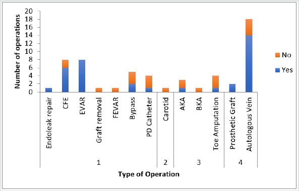
Figure 1: First audit cycle: antibiotics prescribed according to guideline for individual operations.
Lupinepublishers-openaccess-Surgery-Casestudies
1= Vascular surgery (Abdominal and lower limb arterial reconstruction)
2 = Carotid endarterectomy
3 = Lower Limb amputation
4 = Upper limb renal access
CFE = Common Femoral Endarterectomy, EVAR = Endovascular Aneurysm Repair, FEVAR = Fenestrated Endovascular Aneurysm Repair, PD = Peritoneal Dialysis, AKA = Above Knee Amputation, BKA = Below Knee Amputation
Of a total of 60 operations, 5% did not correlate to a category within the guideline (n=3). Of the 57 that did, 63% received antibiotics according to NHS Grampian guidelines (n=36), 21% were prescribed antibiotics that were different (n=12) and 16% did not receive antibiotics (n=9). Figure 1 demonstrates a breakdown of individual operation categories according to whether or not the antibiotics prescribed adhered to guideline (Figure 1).

Different Antibiotics

There were 12 cases that had antibiotics prescribed that were different from the guideline. Of these, 58% were not compliant due to the avoidance in prescribing gentamicin (n=7), 25% were prescribed benzypenicillin and gentamicin as per guideline but without the addition of flucloxacillin (n=3) and 8% missed out appropriate Gram-negative cover (n=1).

Timing of Antibiotics

Of those operations that received antibiotics, 79% were administrated ‘At induction, ≤60 minutes before incision’ as per guideline (n=37), 19% of antibiotics were given too late (n=9) and 2% were given too early (n=1).

Second Audit Cycle

Operations

A total of 28% of all operations did not fit a specific category (n=18). This included a biopsy of a foot ulcer, closure of a fasciotomy, vein operations, antecubital fossa wound debridement, cervical rib resections, thigh loop access graft, incision and drainage of a brachiocephalic fistula abscess and an excision of a prosthetic graft.

Antibiotics Compared to Guideline

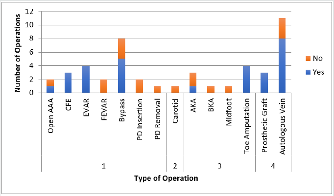
Of the 64 operations, 28% had no antibiotic guideline (n=18). Of the 46 that did, 63% received antibiotics according to guideline (n=29), 28% had different antibiotics prescribed (n=13) and 9% did not receive antibiotics (n=4). Figure 2 demonstrates individual operations and their correlation of antibiotics prescribed according to guideline (Figure 2).
Figure 2: Second audit cycle: antibiotics prescribed according to guideline for individual operation (For abbreviations see Figure 1).
AAA = Aortic Aneurysm Repair
Lupinepublishers-openaccess-Surgery-Casestudies

Different Antibiotics

In total, 12 cases had antibiotics prescribed that were different from guideline. A total of 58% were not compliant due to the avoidance in prescribing gentamicin (n=7), 17% were prescribed benzypenicillin instead of flucloxacillin (n=2), 8% missed out the addition of flucloxacillin (n=1), 8% did not have appropriate Grampositive cover (n=1) and 8% gave gentamicin when it was not needed (n=1).

Timing of Antibiotics

A total of 88% of all antibiotics prescribed were administrated according to guideline (n=30), 9% of antibiotics were given too late (n=3) and 3% of antibiotics were given too early (n=1).

Discusión

The audit reveals important data determining whether local guidelines for antibiotic prophylaxis, during vascular surgery, are being accurately followed. The Getting It Right First Time (GIRFT) is a national programme focused at reducing unwarranted variations and ultimately improving patient care [6].Through the use of local and national collaboration medical professionals are able to analyse results and improve service management. This process is further strengthened by assessing local guidelines and implementing change. Thus, the results of this audit come at a time when post-operative infection rates are at the forefront of the public and governmental interest.

Data Recording

Accurate documentation is key not only for patient safety but also for correct patient management. NHS England has revealed their ‘Five Year Forward View’, which outlines the national target that all documentation be electronic by 2020 [7]. This is intended to improve communication between health care providers, allow for ‘real-time digital information on a person’s health and care’, reduce cost and improve patient safety [8]. During the first audit cycle, 15% of all anaesthetic records and 48% of all operation notes were not uploaded to the electronic system. These initial results are concerning as accurate documentation enables medical practitioners to record complications making future interactions safer. Additionally, with the current ageing population and demands upon healthcare, clinicians are constantly under pressure to reduce the ever-expanding waiting times. This is often achieved with postoperative appointments carried out by different clinicians and more junior staff. Therefore, it is important that all records be easily accessible. These results were highlighted to clinicians and the second audit cycle saw a vast improvement: only 5% of anaesthetic records and 17% of operation notes were not uploaded.

Deviation from Protocol

Another area highlighted by the audit was the lack of documentation regarding the reasons for not using appropriate guidelines. The GMC states that: ‘Clinical records should include relevant clinical findings, the decisions made and actions agreed, and who is making the decisions and agreeing the actions, the information given to patients, any drugs prescribed or other investigation or treatment, who is making the record and when [9]. Thus, decisions regarding differing antibiotic prescriptions require accurate documentation. This allows clinicians to realise that the change from protocol is intended and enables others to understand the rationale behind this. The first audit cycle revealed that 21% of antibiotics were prescribed against protocol with 16% of operations receiving no antibiotic cover. Despite clinician awareness of the current NHS Grampian guidelines increasing throughout the audit intervention, the second cycle revealed similar figures with 28% of antibiotics being prescribing against protocol and 9% of all operations not receiving antibiotics.
The insertion and removal of peritoneal dialysis catheters was a common area of wrongful prescribing. Literature strongly links the insertion of peritoneal dialysis without antibiotic prophylaxis with an increased risk of peritonitis [10]. The International Society of Peritoneal Dialysis recommends in their guidelines the use of antibiotic prophylaxis in peritoneal access with strong evidence for vancomycin [11]. No guidelines exist for the removal of a peritoneal dialysis catheter and antibiotic prophylaxis. This recommendation should be considered when updating the current guideline with a separate section added for peritoneal dialysis. Another area that saw a deviation from protocol was when individuals prescribed either benzylpenicillin instead of flucloxacillin or missed out the addition of flucloxacillin when benzylpenicillin was being used. It is unclear why this was the case as accurate documentation was scarce. One could assume that a lack of understanding regarding the differing organisms covered by each antibiotic could play a role. Thus, further investigation into why this occurred and education into why these antibiotics are included in the current guideline should occur.

Gentamicin

Concerns raised by both the anaesthetic and vascular team included the reluctance to use nephrotoxic antibiotics in procedures associated with high renal injury, such as, those undergoing contrast or requiring clamping of renal vessels. In the current guideline the first- and second-line antibiotic for abdominal and lower limb arterial reconstruction includes gentamicin, a well-known nephrotoxic. 58% of non-compliance in both the first and second cycle was due to the omission of gentamicin. In these cases, individuals were either prescribed teicoplanin alone or metronidazole instead. Single-dose prophylactic gentamicin has been extensively researched with many articles reporting its safety, as toxicity is associated more with therapeutic level duration rather than peak levels.10 However, a study by Nielsen et al. found an increased incidence of acute kidney injury in patients receiving single-dose prophylactic gentamicin during cardiac surgery, but no greater increase in postoperative dialysis or mortality [12]. When considering the administration of gentamicin clinicians must remember the risk factors for its toxicity: older age, reduced renal function, dehydration and concomitant use of diuretics or iodide contrast media [13]. Importantly, these risk factors are present in a substantial proportion of vascular patients. Interestingly, in the NHS Lanarkshire ‘Antibiotic Prophylaxis in Vascular Surgery’ guideline, clinicians are asked to consider reducing the dose of gentamicin from 80mg to 40mg or omitting altogether in patients who are at risk of developing an acute kidney injury [14]. This poses several questions: is the current 120mg dose of gentamicin required or is a smaller dose as effective; should we reduce the dose in patients at risk of an acute kidney injury and could other antibiotics with less nephrotoxicity be considered?

Reclassification as Angiogram Procedures

The risk of surgical site infection (SSI) depends on the type of operation and site. Open procedures carry a greater risk of SSI due to the larger wound created and the exposure of viscera compared to those carried out via angiogram. Skin commensals differ greatly for vascular interventional radiology procedures such as digital subtraction angiograms. Patients within NHS Grampian are most commonly prescribed flucloxacillin and guidelines are currently being developed for these procedures. The argument remains whether patients undergoing Endovascular Aneurysm Repair (EVAR) should be classified according to these interventional guidelines rather than as abdominal and lower limb arterial reconstruction surgery due to the lack of abdominal viscera exposure, thus avoiding the use of nephrotoxics.

Antibiotic Timing

The timing of antibiotics is important to guarantee efficacy and reduce SSI. The optimal administration of antibiotics is within 60 minutes prior to skin incision with the odds of SSI rising significantly out with this time frame [3]. The correct timing of antibiotic administration improved from 79% within the first audit cycle to 88% in the second.

Categorisation

In the first audit cycle 5% of operations could not be categorised compared to 28% of operations within the second. Anterior cervical rib resection for thoracic outlet obstruction is a procedure commonly carried out in ARI, however, this procedure has not been categorised. Similarly, the treatment of varicose veins is commonly carried out and this too fails to be categorised. Although it is not possible to ensure that every procedure fits into an operation category it is important that common operations are grouped so that appropriate antibiotic prophylaxis can be prescribed.

Conclusion

This audit of antibiotics within intraoperative vascular surgery demonstrates interesting results. A clear improvement has been made in data recording, antibiotic timing and antibiotic coverage. However, the results show no clear difference in antibiotics prescribed according to guideline. This could be explained, in part, by the reluctance of clinicians to prescribe nephrotoxic antibiotics as well as the lack of clear categorization of certain procedures. Thus, an update of the current guideline is required to improve antibiotic prescription and patient care. In an era of Antibiotic Guardianship, it is imperative in order to reduce morbidity and mortality that appropriate antibiotic prescribing is adhered to and practitioners do not become complacent.

For more Lupine Publishers Open Access Journals Please visit our website:
For more Surgery Journal articles Please Click Here:
To Know More About Open Access Publishers Please Click on Lupine Publishers

Follow on Linkedin : https://www.linkedin.com/company/lupinepublishers
Follow on Twitter   :  https://twitter.com/lupine_online

Friday, October 25, 2019

Lupine Pubishers| Cervical Spinal Cord Injury During Coronary Artery Bypass Graft Surgery in An Elderly: A Case Report

Lupine Publishers | Journal of Surgery & Case Studies


Case Report


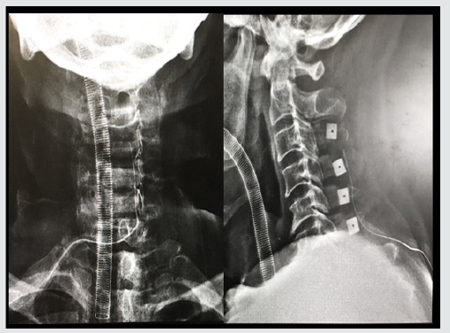
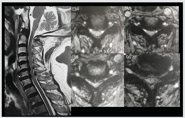
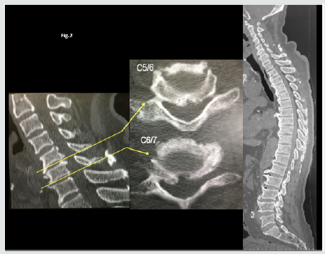
Intraoperative spinal cord injury (SCI) is uncommon and the pathology has not cleared. We report cervical cord injury without vertebral fracture during a coronary artery bypass operation. Cervical cord compression (CCC) is compression of the cervical spinal cord that occurs during the normal course of aging and may progress into cervical spondylotic myelopathy (CSM), which can cause neurologic dysfunction. Many people with cervical cord compression are asymptomatic. However, patients with CCC are at higher risk of SCI following minor injury. An 85-year-old woman with asymptomatic cervical cord compression underwent a coronary artery bypass graft surgery. After the surgery, he developed tetraplegia. MRI revealed severe spinal cord compression from C3 to C7 and C5/6 signal change of spinal cord (Figure 1). The radiograph and CT showed diffuse idiopathic skeletal hyperostosis and thoracic kyphosis of 55 degree (Figure 2). Cervical SCI after was diagnosed. After the diagnosis, we underwent laminoplasty (Figure 3). And he almost completely recovered after surgery.
Figure 1: Magnetic resonance imaging (MRI) of the cervical spine.
Lupinepublishers-openaccess-Surgery-Casestudies
Figure 2: MRI showed multilevel spinal cord compression.
Lupinepublishers-openaccess-Surgery-Casestudies
Figure 3: The radiograph and CT showed diffuse idiopathic skeletal hyperostosis and thoracic kyphosis Radiograph after laminoplasty for cervical spinal cord injury.
Lupinepublishers-openaccess-Surgery-Casestudies
Iatrogenic cervical SCI after non spinal surgeries that requires neck hyperextension is rarely reported, probably due to underdiagnosis and underreport. In the current aging society, the prevalence of patients with CCC is expected to increase. In our population-based magnetic resonance imaging (MRI) study, the prevalence rate of CCC was 24.4%, and it was higher with increasing age in both men and women [1]. Among the 15 cases (including ours) published in the literature [2], most patients had cervical cord compression and were old men. Eight patients had undergone coronary artery bypass surgery that requires prolonged operative time. Only 3 patients had almost complete recovery. Two patients required tracheostomy for long-term ventilator support. Two patients died. These cases reiterate the potential risk of iatrogenic SCI in people with predisposing conditions such as cervical spondylosis and CCC. Moreover, in this case, the patient had diffuse idiopathic skeletal hyperostosis and thoracic kyphosis of 55 degree, which can lead inflexibility of spine. The inflexibility of thoracic spine might cause hyperextension of cervical spine during the surgery. Surgeries requiring prolonged neck hyperextension put patients with cervical spondylosis and CCC at risk for SCI. It is essential to recognize the potential occurrence of iatrogenic SCI might endanger patients’ lives.

For more Lupine Publishers Open Access Journals Please visit our website:
For more Surgery Journal articles Please Click Here:
To Know More About Open Access Publishers Please Click on Lupine Publishers

Follow on Linkedin : https://www.linkedin.com/company/lupinepublishers
Follow on Twitter   :  https://twitter.com/lupine_online
Saturday, October 12, 2019

Lupine Publishers | All-On-Four Treatment Concept in Dental Implants: A Review Articles

Lupine Publishers | Journal of Surgery & Case Studies


Abstract


Edentulism has been demonstrated to have negative social and psychological effects on individuals that include adverse impacts on facial and oral esthetics, masticatory function and speech abilities, that when combined, are translated into significant reductions in patients’ quality of lives. It is well-known that immediate placement of implants is a challenging surgical procedure that requires proper treatment planning and surgical techniques. There are several prosthetic options to rehabilitate severely atrophic maxillae and mandibles have been developed such as conventional complete dentures, implant supported removable and implant supported fixed prosthesis. Implant supported prosthesis may not be feasible in many conditions because of the vicinity of vital anatomical structures, poor bone quality and quantity.
The implant technology developed as the all-on-four concept is offered as an alternative to conventional implant applications. In the all-on-four technique, 4 implants are placed in the inter foraminal region in the mandible and in the pre-maxillary region in total edentulism cases. An exception is the all-on- Four concept, which uses only 4 implants to support an acrylic, screw-retained provisional prosthesis delivered on the day of implant placement, followed by a definitive prosthesis approximately 4 months later. The patient can also be benefited with the immediate rehabilitation of edentulous arches which helps maintaining self-confidence and quality of life.
Keywords: All on four concept; Dental implant; Tilted implant; Severe atrophy
Abbreviation: CT: Computerized Tomography; Ncm: Newton centimeter ; mm: Millimeter

Introduction

One of the greatest challenges in implant dentistry is the treatment of patients with severely atrophic jaws. Such atrophy can be horizontal or vertical or both; even if sufficient vertical bone is present, lack of ridge width can still preclude treatment with implants that are 4 mm in diameter or wider. Over the years, many techniques, procedures, and materials have been introduced to solve the complex problems associated with treatment of atrophic jaws. One early approach in the posterior maxilla was to augment the sinus in conjunction with the simultaneous placement of implants [1–3]. In cases where the mandibular bone height is less than 12.0 mm, tissue augmenting techniques including bone ridge expansion and bone grafts are typically made necessary to allow the placement of traditional dental implants. Despite their widespread use, these techniques are clinically very challenging and display higher levels of tissue and implant morbidity.
Modern oral rehabilitation strategies based on the utilization of dental implants and immediate loading techniques, have been demonstrated to allow for the rapid and efficient restoration of patients with superior esthetic and masticatory properties, while reducing the morbidity and damage to both soft and hard tissues. In some cases of the completely edentulous patients, implant supported prosthesis treatment is almost impossible without complex techniques such as nerve transposition and grafting in the posterior maxilla and mandible. A solution for such situations is the All-on-4 concept [4]. Implant supported prosthesis may not be feasible in many conditions because of the vicinity of vital anatomical structures like mandibular canal or maxillary sinus. Over the years, many techniques, procedures, and materials have been introduced to solve the complex problems associated with treatment of atrophic jaws and avoid the vicinity of vital anatomical structures such as the use of short implants, alveolar distraction osteogenesis, guided bone generation, use of intraoral and extraoral autogenous bone grafts, nerve repositioning etc. The high cost, time and morbidity associated with such approaches have limited their application.
At the beginning of the millennium, distally tilted implants were proposed, enabling the use of denser bone located in the anterior mandible for improving bone anchorage, and replacement of posterior teeth without extended cantilevers and avoiding bone grafting procedure [5]. The “all-on-four” treatment concept was developed to maximize the use of available remnant bone in atrophic jaws, allowing immediate function and avoiding regenerative procedures that increase the treatment costs and patient morbidity, as well as the complications inherent to these procedures [6].

History of the All-on-4® concept

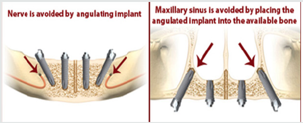
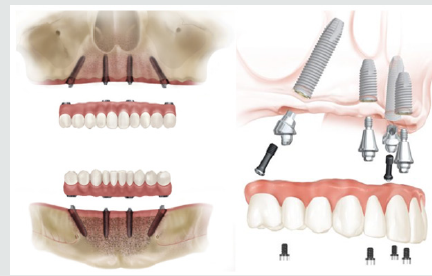
One of the early designs of the All-on-4 style concept can be traced back to Mattson and colleagues, in 1999 whereby they treated, patients with severely resorbed edentulous maxilla by inserting 4 to 6 implants in the premaxilla to avoid sinus augmentation and successfully restored them with fixed prosthesis with 12 teeth supported by superstructure [7]. In 2000, Krekmanov and colleagues were also able to demonstrate posterior tilted implantsupported prosthesis was possible. The All-on-4 immediate loading concept was developed, institutionalized and systematically analyzed in the 2003 by a dentist Paulo Malo and colleagues [8]. This concept uses 2 vertical anterior implants in conjunction with 2 distally tilted inclined implants with their apices positioned anterior to the sinus wall or mental foramen (Figure 1). It involves the use of straight and angled multiunit abutments, which support a provisional, fixed, and immediately loaded, full arch prosthesis. It has been developed to maximize the use of available bone and allows immediate function [9] (Figure 2).
Figure 1: Show how vital structure avoided in All on 4 Concept.
Lupinepublishers-openaccess-Surgery-Casestudies
Figure 2: Show All on 4 concept design and multi-unit abutment.
Lupinepublishers-openaccess-Surgery-Casestudies
In this technique, four implants are placed in the anterior region of the jaw between the two mental foramina in the mandible and between the mesial walls of the maxillary sinus in the maxilla. The two anterior implants follow the jaw anatomy and the two distal implants are tilted at 45° angulation (Figure 1). Many longterm studies and published data on the All-on-4 concept reported cumulative survival rates between 92.2% and 100% [10].

Treatment Planning

The All-on-4 clinical solution has been developed to
a. Maximize the use of available bone
b. To allow for Immediate Function.

General Considerations

a. Ability to achieve primary implant stability (35 – 45 Ncm)
b. No severe parafunctions
c. To diminish the cantilever, tilt the posterior implants.
d. Does not require a wider opening of the mouth
e. It is advisable to place implants between extraction sockets.
f. Indicated with a minimum bone width of 5mm and minimum
bone height of 10mm from canine to canine in maxilla and 8mm in mandible. g. If angulation is 300 or more, the tilted implants can be splinted. For tilted posterior implants, the distal screw access holes should be located at the occlusal face of the first molar, the second premolar, or the first premolar.

Advantages of the All-on-4 Concept

I. Avoids complex surgery,
II. Less invasive procedure for the pt.
III. Graft less procedure.
IV. Implants well-spaced, good biomechanics, easier to clean, immediate function and aesthetics
V. Simplified surgical & prosthetic procedure.
VI. Reduced cost due to less number of implants.
VII. High success rates.
VIII. Angled posterior implants:-
a. Avoid anatomical structures
b. Allow longer implants anchored in better quality bone
c. Reduces posterior cantilever

Disadvantages

a. Length of cantilever in the prosthesis cannot be extended beyond the limit.
b. Free hand arbitrary surgical placement of implant is not always possible as implant placement is completely prosthetically driven.
c. It is very technique sensitive and requires elaborate presurgical preparation such as CAD/CAM, surgical splint [11].

Treatment Protocol Consists of Two Phases: Surgical and Prosthetic

I. Surgical Protocol
II. Prosthetic Phase
I. Step 1: Selection of case satisfying the inclusion criteria
II. Step 2: Planning implant placement using All-On-4 Guide (Prefered)
III. Step 3: Location of Maxillary Antrum and Mental Foramen with All-On-4 Guide
IV. Step 4: Implant placement done following the protocols
The surgical guide is placed into a 2 mm osteotomy that is made in the midline position of the maxilla or mandible and the titanium band is contoured to follow the arc of the opposing arch. The mandibular guide also assists in retracting the tongue. The vertical lines on the guide are used as a reference for drilling at the correct angulation, which should not be greater than 45º. Angulated pins and denture can also be used as templates. The two most anterior implants follow jaw anatomy in direction (lingual tilting in cases of severe mandibular resorption). The two posterior implants are inserted just anterior to the foramina or maxillary sinus and tilted distally approximately 30°– 45° relative to the occlusal plane. Implant torque is set to greater than 35 Ncm. If the torque values for three or more implants are below 35 Ncm, a 2-stage technique is preferred [9].

Prosthetic Phase

17º multiunit abutments and 30º angulated, Straight Abutments with different collar heights are placed onto the implants. These are used to achieve the correct access allowing relative parallelism and so that the rigid prosthesis can be seated passively [12]. For provisional prosthesis fabrication on the day of surgery (2–3 hours after surgery), the impression copings are placed onto the multiunit abutments which are then splinted with autopolymerising resin and wire bars. It ensures an accurate transfer without accidental displacement of the impression copings and impression is made. Protective healing caps are placed on the abutments while provisional is being made. Acrylic provisional screw retained prosthesis is torqued to 15 Ncm. Only soft diet is recommended. For final prosthesis (4–6 months after initial implant placement), if the implants are judged stable, provisional restoration is removed and bite is registered. Multiunit laboratory analogs are attached to the provisional and then it is mounted on an articulator against a counter model. The prosthesis is indexed with putty. Resin pattern is fabricated in sections and the sections are joined in the patient’s mouth. This resin pattern gets scanned and framework is made by CAD/CAM technology. After try-in of the framework, final prosthesis is fabricated and delivered. Final prosthesis can be a metal-acrylic resin with a titanium framework and acrylic resin prosthetic teeth, or a metal ceramic prosthesis with titanium framework and all ceramic zirconia crowns [12].

Discusión

A recent shift in practice paradigm has been to minimize treatment costs and patient morbidity while providing the most satisfying patient-centered treatment outcomes according to the state of the art of dental practice. The “All on-Four” treatment concept is an attempt to reach these objectives by providing relatively straight forward, predictable treatment option to rehabilitate edentulous patients with a high outcome of Quality of life [13]. Babbush et al. described a protocol of combining the 3.5-mm-diameter Nobel Active implants with the All-on-4 concept and reported on the results of 227 implants after 1 to 3 years of Follow-up. The cumulative survival rate was 98.7% at the end of 3 years, with a 100% prosthetic survival rate [14]. Paul Malo did retrospective clinical studies and found that long-term outcomes ( up to 10 years ) of 245 patients with completely edentulous mandibles rehabilitated with the All-on-4 treatment concept and reported cumulative implant survival rates of 93.8% and 94.8% using the patient and implant as units of analysis, respectively [15] Lopes et al. did a prospective study to evaluate the medium- and long-term outcomes of the rehabilitation of completely edentulous jaws in immediate function with the All-on-4 treatment concept using a computer-guided surgical protocol and reported an overall 96.6% cumulative survival rate after 5 years of follow-up [16]. Published studies on the All on-Four concept have shown cumulative survival rates to range between 92.2% and l00% [10]. clinical research over the past decade has shown that more than 4 dental implants to support a full, fixed (non-removable) bridge is typically not necessary, and that placing more than 4 implants does not increase success rates[17]. Owing to the freedom of tilting, the implants can be anchored in dense bone structures (anterior bone with higher density) and well spread anteriorly-posteriorly giving an effective prosthetic base. By reducing the number of implants to four, each implant can be placed without coming into conflict with adjacent implants. This treatment approach, using tilting and few implants rather than inserting several implants competing for space, has demonstrated good results.

Conclusion

The “All-on-4” treatment concept seems to be an alternative option for rehabilitating edentulous jaws compared with advanced surgical approaches without using removable prostheses. It is a cost-effective procedure, decreasing the treatment times, the morbidity and allowing a higher patient quality of life. Placement of dental implants previously in attempts to treat the severely resorbed maxilla and mandible has had only limited success. But the rehabilitation of completely edentulous, atrophied maxilla and mandible by the placement of implants using the AII-on-Four protocol gives new hope for a perceivable success, while becoming a promising treatment method of choice and standard in the care for severely compromised patients.

For more Lupine Publishers Open Access Journals Please visit our website:
For more Surgery Journal articles Please Click Here:
To Know More About Open Access Publishers Please Click on Lupine Publishers


Friday, September 27, 2019

Lupine Publishers | Postoperative Biliary Peritonitis

Lupine Publishers | Journal of Surgery & Case Studies


Annotation


The author watched 83 patients with postoperative peritonitis bilious, of whom 27 (32.5%) the reason for its development was inspired by the ongoing peritonitis, 38 (45.9%) is non-suite Lodge of the gall bladder, 3 (3.6%)-damage to abnormally developed bile ducts, 8 (9.6%)-migration tubes exegesis, 7 (8.4%)-wound exegesis. Died 8 (9.6%) patients. The author notes the difficulty of diagnosis of this complication due to the paucity of symptoms.

Keywords:Operation; complication; bile peritonitis; diagnosis; treatment

Introduction

Lately there has been a significant increase in patients with acute cholecystitis, if this fatality rate when it is 3-7% [1]. Analysis of literature data suggests that even now, despite great technical capabilities of modern surgery, surgeries for cholecystitis are accompanied by various tactical and technical medical errors. So, if damage reaches exegesis cholecystectomy 5.6% and zhelcheistechenie from gallbladder bed is 8.3% [2]. In 68%- 81.1% of cases the reason for the development of postoperative biliary peritonitis are tactical and technical medical errors [3,4,5]. Prominent negative role for prediction of treatment provides and the presence of patients expressed disorders of immunity and slowing the regeneration process [5]. The most frequent complications arise during surgery in patients with destructive form of this disease, the syndrome of Mirizzi, sclerosis of the gallbladder and biliary tract abnormalities. Postoperative mortality in persons aged patients reaches 80% [2.5]. Prognosis worsens or if there already or developing in the patient of acute pancreatitis. Such observations lethality reaches 30-35% [1]. These data demonstrate the feasibility of evaluating the treatment of patients with acute cholecystitis in the various hospitals to develop a diagnostic and treatment algorithm specified complications.

Material and Methods

The OCG in surgical units were MUNICIPAL treatment 5412 patients with acute cholecystitis. Men (33.7%), 1828 and women-3584 (66.3%). the age of the patients was from 21 until 95 years. Operations are performed at 4106 (75.8%) patients, among them a laparoscopic way-from 1728 (42.1%), conversion option-250 (6.1%) Postoperative lethality was 2.18%. All patients, including those under the age of 50 years, revealed on 2 or more For Co morbidities. Emergency operations were performed at 2464 (60%) patients. Destructive form of inflammation identified at 3116 (75.9%) patients and it has always been associated with the more or less pronounced inflammatory infiltrate around the gallbladder with involvement in the pathological process of adjacent liver tissue and liver the duodenum bundles, with 507 (14.8%) He was a development density. Out of the total number of cholecystectomy (4106) postoperative biliary peritonitis, varying degrees of prevalence was observed in 83 (2.02%) patients. At 8 (0.19%) of them he was the cause of death.
A complication they had revealed before relaparotomy and confirmed during the repeated surgical intervention. For the diagnosis of this complication conducted clinical and laboratory, biochemical and instrumental examinations, including ultrasound, CT scan, x-ray and laparoscopy, abdominal and thoracic cavities. From 83 patients were carried out at relaparotomy 22 (26.5%) of them have 18 with symptoms of widespread peritonitis and 4 after random enforced pulling the drain tube from the patient when turning-exegesis nabob (among these patients during relaparotomy discovered a clump of bile under the liver). At 41 (49.4%) same patient relaparotomy was replaced by venting bile accumulation zones (in the right podreberie, right side canal and pelvic cavity) under ultrasound control. Reasons for the development of postoperative bile peritonitis were: continuing peritonitis-27 (32.5%), non-suite lodge gallbladder-38 (45.9%), corruption of abnormally developed bile ducts-3 (3.6%), the migration of the drainage tube from exeresis-8 (9.6%), wound choledochitis-7 (8.4%) On clinical flow disease from 83 patients, only 12 (14.4%) There were symptoms of peritonitis, and 71 (85.6%)-they were erased.
On the prevalence of abdominal lesion bed (limited and unlimited) peritonitis diagnosed at 65 (78.3%), and common-u 18 (21.7%) should indicate if local peritonitis symptoms of peritoneal sepsis was observed then when they were distributed in all patients, and 5 (6%) He even accompanied the infectious-toxic shock (these patients died). Marked by a certain relationship between severity of endogenous intoxication and volume zhelcheistechenija. So, if you lose your 300-500 ml of bile develops local peritonitis with mild degree of intoxication, 500-1000 ml-diffusive-spilled with average and more than 1000 ml-General severe peritonitis. From 83 patients with Leukocytosis, with a shift of Leukocyte formula left, there were only 31 (37.3%). Thus, in the majority of patients leading diagnostic test was abundant expiration of bile from the abdominal cavity, or drainage, or past them.

Results

5412 of patients with acute cholecystitis (1.66%) and 90 died people from postoperative bile peritonitis 8 (0.16%). The remaining 82 patients died from other causes. From 83 patients with postoperative peritonitis bilious died 8 (9.6%) of the 27 patients with peritonitis was before surgery and he continued after she died 4 (15%), conditionally from 38 patients with nonushitym gallbladder bed-1 (2.6%) From 3 patients where the damage occurred an abnormally developed bile ducts, there have been no deaths. Of the 8 patients who died of the drainage tube migration, died 1 (12.5%), conditionally of 7 patients with wound choledochitis died 2 (28.5%) of probation. We believe it necessary to point out that damage to abnormally developed bile ducts and was immediately identified exeresis wound during the execution of the primary operation and steps were taken to restore the tightness of biliary tract, bile peritonitis but evolved, Despite the timely diagnosis of this complication. All 10 patients were performed relaparotomy during which only 8 have external drainage of biliary tract, and the 2-x stitches on holedoh, but after 1 day they prorezalis, accompanied by a progression of his fatal peritonitis exodus.

Discusión

Thus, the development of postoperative bile peritonitis was due to tactical and technical medical mistakes made as when choosing how to gall bladder removal and manipulation on this body and its surrounding tissues, and also when draining the abdominal cavity. A negative role played and the older patient’s violations of immunity. If you get the drain tube from the exegesis, diagnose the development of this complication did not pose any difficulty, when his other reasons it was difficult-because of wear of clinical symptoms. This can be attributed to the conduct of postoperative corrective medical therapy, use of antibiotics and pain medication. However, the patients symptoms of intoxication (tachycardia and dehydration) and euphoria, but the main thing-the expiration of bile by drainage tubes, allowed or suspect the complications, or conclude the progression of peritonitis if he had before the surgery. Severity of the pathological process is largely dependent on zhelcheistechenija. So the lowest mortality was observed in patients with defects in the closing of the lodge of the gall bladder.
It is possible to explain the cover bed great seal and other soft tissues, limiting the speed of zhelcheistechenija. Rate and amount of bile in the free abdomen mostly depend on the diameter and the nature of the damage to the bile duct, as well as on the availability of occlusion is below the level of his trauma. The greater was hypertension, the faster evolved intoxication, with 8 (9.6%) patient’s zhelcheistechenie to drain the tubes should not exceed 50 ml, and took place by them. Effaced symptomatology was the main reason the diagnosis on complications lag 1-2 days. With the increase in the number of zhelcheistechenija is progressing not only peritonitis, but endogenous toxemia. In such cases, the expectant surgical treatment becomes dangerous to the life of the patient. However, the mere relaparatomy or drainage of abdominal cavity under ultrasound control does not ensure success in the treatment of this complication. To do this, you must use complex medication therapy, which helps secure the correction vodno-elektrolitnogo balance, renal and hepatic failure to conduct the fight against toxemias and microbial aggression.

Conclusion

Postoperative biliary peritonitis usually driven by technical and tactical mistakes when performing cholecystectomy. Most frequently surgeons admit negligence when closing a lodge of the gall bladder. Rough pricking the surrounding liver tissue accompanied by damage to needle close located vnutripechenerngo bile duct that is accompanied by the expiration of bile into the abdominal cavity-free. In such cases rarely imposed the seams do not provide impermeability Lodge. Risk of bile peritonitis increases dramatically when injury anomalous biliar no traumatic ducts. For this reason, when determining the patient’s development anomalies Brigade must immediately be turned surgeon, has extensive experience in biliary tract surgery. Particular nuisance calls cause gall development of peritonitis such as migration of the tube, which the surgeon introduced clearance exegeses. Most often this occurs when the patient is rotated on its side. However, it tube prolapsed may occur and when the surgeon does not take into account its length from exegeses and up to the abdominal wall.
If it is short, when awakening the patient strains, inflates, twitches, and as a result dramatically increases the distance from exegesis and to the anterior abdominal wall and tube vydjorgivaetsja of duct, that is to create a vnutribrjushinnyj supply of its length. Wound choledochitis usually occurs when a patient has a dense inflammatory infiltrate. Of particular concern is the incidence of severe biliary peritonitis while destructive-nekroticheskom lesions of gallbladder and pancreas. These patients require not only adequate drainage of the abdominal cavity, but also sound therapy and antifermentnuju is pathogenetically local hypothermia pancreas.

For more Lupine Publishers Open Access Journals Please visit our website:
For more Surgery Journal articles Please Click Here:
To Know More About Open Access Publishers Please Click on Lupine Publishers


Saturday, September 14, 2019

Lupine Publishers | 56 Years of Experience of Primary Tumors of The Heart

Lupine Publishers | Journal of Surgery & Case Studies


Abstract


From 1963 to 2019 7,600 open Heart operations were done in ouers diferents centers in Argentina. 23 Patients (0.3%) were operated on of primary Tumor of the heart. In this report, we review the surgical experience and results of cardiac tumors during this 56 years Material and methods 15 patients were female (65%) The average age was 52 years (1967) The clinical presentation was Dyspnea 8 (34,8%) Arrtymia 6 (26,08%) ChestPain 3 (13%), Emboli 2 (8,7%), Syncope 2 (8,7%), Accidentaly 2(8,7%). 17 were Myxomas 17 cases (74%) 15 (88%) left and 2 (12%) rigth. Papillary Fibroelastoma 4 (17,4%) sarcoma 1 (4,4%) Cyst 1 (4,4%) Surgical Thecnique: all of the patients in this serie were operated on with Cardiopulmonary Byapass Results The operative mortality was 1 case (4,3%). The patient with a Sarcoma died inmediatly after the operation; the entire Rigth Ventricle was invaded by the Tumor the exsicion was very dificult. The rest of the patients survived the operation At the average of 29, 8 years of follow up of the 17 patients with Myxmomas 6 (35%) died ,no recurrence of the Tumor were found in this patients. In the patients with Papillary Fibroelastoma 2 patients were lost of follow up one died at 12 years after de operation and the other is alive The patient withCyst died at 8 years after the operation conclusión Most of the primary Tumors of the Heart have good Surgical Prognosis is vey important the early detention.
Keywords:Primary Tumors of the Heart; Surgical of Tumors of the Hearts; Surgery Tumors of the Heart

Introduction


Primary cardiac tumors can be classified as either benign/ malignant or primary/secondary of malignant pathology. Data from 22 large autopsy series reported by Mc Allister et al [1]. showed the frequency of primary cardiac tumors is ∼0.02%, corresponding to 200 tumors in 1 million autopsies. Around 75% are benign; nearly half are myxomas; and a majority of the rest are lipomas, papillary fibroelastomas, and rhabdomyomas [1]. Sixty years ago in 1954, Crafoord [2] was the first to excise an atrial myxoma on cardiopulmonary bypass [2]. Today, cardiac tumors represent around 0.3% of all open-heart surgeries [3]. From 1963 to 2019 7,600 open Heart operations were done in ouers diferents centers in Argentina. 23 Patients (0.3%) were operated on of primary Tumor of the heart. In this report, we review the surgical experience and results of cardiac tumors during this 56 years.

Material and Methods


15 patients were female (65%) The average age was 52 years (1967) The clinical presentation was Dyspnea 8 (34,8%) Arrtymia 6 (26,08%) ChestPain 3 (13%), Emboli 2 (8,7%), Syncope 2 (8,7%), Accidentaly 2(8,7%). 17 were Myxomas 17 cases (74%) 15 (88%) left and 2 (12%) rigth. Papillary Fibroelastoma 4 (17,4%) sarcoma 1 (4,4%) Cyst 1(4,4%) Surgical Thecnique: all of the patients in this serie were operated on with Cardiopulmonary Byapass 21 througth Sternotomy and 2 Minimally invasive Rigth Thoracotomy. In 20 patients the tumor was excised under direct visión with and without magnification. In 3 patients Video 3 D was used to improved the visualization during the operation.

Results


The operative mortality was 1 case (4,3%). The patient with a Sarcoma died inmediatly after the operation; the entire Rigth Ventricle was invaded by the Tumor the exsicion was very dificult. The rest of the patients survived the operation At the average of 29,8 years of follow up of the 17 patients with Myxmomas 6 (35%) died ,no recurrence of the Tumor were found in this patients. In the patients with Papillary Fibroelastoma 2 patients were lost of follow up one died at 12 years after de operation and the other is alive The patient withCyst died at 8 years after the operation.

Discusión


The most common site of attachment of the myxoma is in the fossa ovalis. Accounts for approximately 70% of all cardiac tumors. Locations of this tumor in the general population are: 75% in the left atrium (LA), 23% in the right atrium (RA), and only 2% in the ventricles [4-6] Other rare arising of CM are the hearts valves Multiple locations present in 50% in familial forms [7-9]. The clinical signs, manifestations, and symptoms produced are non-specific and determined by the location, size, and mobility of tumor. The main patterns of clinical presentation for patients with myxoma are in general in all the experiences; dyspnea, syncoptic episodes, arrhythmia, palpitations, congestive heart failure, and sudden death, systemic embolism) In ouer experience w the most común clinical presentation was Dyspnea follow by Arrtymia. After clinicopathologic analysis of 61 patients with this tumor were concluded that pathological profiles of cardiac myxoma are not related with the clinical presentation [10].
When diagnosis has been established the treatment of choice for this tumor is surgical resection and in most cases it is curative. The median sternotomy, cardiopulmonary bypass, aorta, and bicaval cannulation with or without cardioplegic heart arrest is the standard approach to resection of these tumor. Novel minimally invasive surgical techniques such as video-assisted or totally robotic resection were described in the literature [11,12]. Surgical approach (atrial or ventricular or aortic) depends from location and size of the mass. While the access of the tumor may be different in most cases, however the general rule to resection of these tumor should be remain. Manipulations of the tumor during resection must be minimizing to prevention peripheral embolization. Complete excision of the mass with the attachment area in the cardiac structures (atrial septum, ventricular septum, atrium or ventricles free wall, and cardiac valves) should be performed. Recurrence may occur within a few months to several years after the initial surgical excision.
The recurrent frequency of myxoma is about 1% to 3% in sporadic forms, 12% in familial forms, and 22% in complex forms. Regular follow-up with clinical examinations and TTE study is recommended in patients with familial CM because of these patients have a significantly higher risk of recurrence. Causes of multiple recurrences include familial forms of the tumor, incomplete excision, intra-cardiac implantation from the original tumor, and malignant transformation. Genetic screening of patients with recurrent cardiac myxomas might help to identify patients at risk for additional recurrence [13,14] Papillary fibroelastoma is a rare benign cardiac tumor (7%–9% of benign primary tumors second after cardiac myxoma as more frequent cardiac valvular tumor [15,16] In 85% of cases, it is a valvular disease affecting aortic valve (35%–63%), mitral valve (9%–35%), tricuspid valve (6%–15%), and pulmonary valve (0.5%–8%) and in a very few cases nonvalvular sites, with left ventricular masses documented on the septum [15,16] and the outflow tract [17,18] Fibroelastomas have also been reported on the right ventricular papillary muscles.
The atrial side of the mitral valve is more often affected by the disease and dimensions may vary from 2 to 70 mm [19] I ouer experience 2 of the tumors were in the Aortic Valve and required to change the valve , one in the mitral valve was exiced without change the valve and another in the ventricular septum Papillary fibroelastoma is a rare, more often incidentally found, form of benign cardiac tumor, Its early diagnosis is paramount to avoid complications such as pulmonary or paradox embolism into systemic circulation. Diagnostic workup to exclude myxomas and valvular endocarditis must be careful. Clinical history, characteristic, size, and position are generally useful for differential diagnosis; echocardiography usually shows a small, mobile, pedunculated, or sessile valvular or endocardial mass, which on many occasions flutters or prolapses into the cardiac chambers during systole or diástole The operation is in general simple and has a good prognosis Cardiac sarcomas usually present with insidious symptoms in young and middle age patients.
They generally possess a poor prognosis with overall survival ranging from 6 to 12 months [20,21] Symptoms related to cardiac sarcomas are variable and may vary from specific cardiac symptoms (pericardial effusions with tamponade, arrhythmias, valvular dysfunction, intracardiac blood flow abnormalities, congestive heart failure, peripheral embolization with systemic deficits, dyspnea, chest pain, syncope, hemoptysis, sudden cardiac death) to general symptoms of neoplastic diseases like fever, malaise and weight loss in general is very dificult to exicesed entire and sometimes require to replace the heart [22-24] A review of the published papers appears to indicate that there are two distinct types. The one type is cystic and is discovered incidentally as a superficially situated lesion lined by ciliated epithelium. The other type is situated in the low interatrial septum or membranous portion of the interventricular septum and has a tubular or trabecular pattern [25] Ouer case was localized in the trcuspid valve and was very easy to excised. In general the Surgical treatment of the most frecuents primary tumors of the heart have good prognosis, is very important the early detection.

Conclusion


Surgical resection of cardiac myxomas contributes in an excellent prognosis and associated with low complications and recurrences rate. Regular long-term follow-up is recommended in all patients with cardiac myxoma particularly in patients with familial form of CM. Familial CM affects the patient’s treatment, follow-up, and his family screening (minimum) with TTE The papilary Fibroelastoma en general is very easy to excised and the Cyst in general also .The surgery of Sarcoma is in general very dificult and many times require an other intervention Most of the primary Tumors of the Heart have good Surgical Prognosis is vey important the early detention.

For more Lupine Publishers Open Access Journals Please visit our website:
For more Surgery Journal articles Please Click Here:
To Know More About Open Access Publishers Please Click on Lupine Publishers

Follow on Linkedin : https://www.linkedin.com/company/lupinepublishers
Follow on Twitter   :  https://twitter.com/lupine_online
 

Gallstone Ileus in the Elderly: Still a Challenge, Report of a Case with Review of the Current Literature

  Abstract Introduction:  Gallstone ileus is described as an intestinal obstruction caused by luminal gallstone impaction. It is a mainly ...