Friday, December 10, 2021

Lupine Publishers | The Case Number 130 of Townes Brocks Syndrome

Lupine Publishers | Surgery & Case Studies: Open Access Journal


Abstract

Townes Brocks syndrome is a very rare genetic syndrome with 129 well-documented patients reported in the medical literature. Townes Brocks syndrome has not been reported before in Iraq. The main aim of this book is to describe the first case of this syndrome in Iraq which seems to be the case number 130.

Introduction

Townes Brocks syndrome is a rare autosomal dominant hereditary disorder which was probably first described in 1972 by Dr Philip L. Townes and Dr Eric Brocks. Dr Philip was professor of pediatrics at the University of Rochester, and Eric Brocks was a medical student. The syndrome is characterized by a triad of imperforate anus, limb defects, and ear abnormalities [1,2].

Main features of the disorder include [1,2]:

a. Anorectal malformations including imperforate anus (absence of an anal opening), recto-vaginal fistula, anal stenosis, unusually placed anus.

b. Hand and foot abnormalities including hypoplastic thumbs, fingerlike thumbs, syndactyly (webbed fingers/toes), fusion of the wrist bones, overlapping foot and/or toe bones.

c. Abnormalities of the ears with sensori-neural or conductive hearing impairment or loss or deafness.

d. Other organ abnormalities including hypoplastic kidneys, multi-cystic kidneys, dysplastic kidneys, and congenital heart defects such as tetralogy of Fallot and defects of the ventricular septum.

The main aim of this book is to describe the first case of this syndrome in Iraq which seems to be the case number 130.

Case report

Figure 1: The girl had low set ears and deformity of the right foot with the presence of only three toes. There was no obvious abnormality of left foot, but the big toe was relatively large.

R.J was first seen at about the age of four months during November 2018 because of poor feeding, failure to thrive, poor response to sounds, and poor head control. The girl also had low set ears, and deformity of the right foot with the presence of only three toes. There was no obvious abnormality of left foot, but the big toe was relatively large (Figure1). She was delivered at 38 weeks by cesarean section. She didn’t pass motion and was found to have imperforated anus. She had colostomy, and the surgeon reported that the sigmoid was not present. The five-centimeter colon ended at the pelvis, and cecum found on the left side. Cloaca treated with diversion colostomy. The parents were relatives and have three normal children. Echocardiography performed during the first month showed normal findings. Brain ultrasound performed on the fifth of August 2018 showed normal findings. Abdominal ultrasound was also performed on the fifth of August 2018 and showed small hypoplastic right kidney (18 x 12 mm) with normal shape. At the age of forty-six days (16, August 2018), a second abdominal ultrasound showed small hypoplastic right kidney. The left kidney had normal size.

Discussion

Authors from Germany, the Netherlands, the UK, the USA, Belgium, Italy , Switzerland and the Czech republic (Jürgen Kohlhase et al ,1998; Jürgen Kohlhase et al ,1999) defined Townes Brocks syndrome as a rare autosomal dominant malformation syndrome with a combination of anal, renal, limb and ear anomalies. Townes Brocks syndrome is a very rare genetic syndrome with 129 welldocumented patients reported in the medical literature [1,2]. In this paper the first case of this syndrome in Iraq is reported which is the case number 130.

Read More Lupine Publishers Surgery and Case Studies Journal Articleshttps://surgery-casestudies-lupine-publishers.blogspot.com/

Friday, December 3, 2021

Lupine Publishers | Multidisciplinary Management of Elderly Cancer Patients: The Radiation Oncologist’s Point of View

 Lupine Publishers | Journal of Surgery and Case Studies


Abstract

Oncological treatments of elderly patients are extremely complex; so far there’s no agreement even on the definition of “geriatric patient”. From the point of view of global health, the problem is of the outmost importance as the number of older patients will increase dramatically in the next years, leading to a change in world epidemiology with a significant increase of chronic-degenerative diseases such as cancer. For this reason, it’s mandatory to provide clinical oncologists with multidisciplinary algorithms aiming at the best treatment of older cancer patients.

Materials and Methods

The complexity of oncological treatments for elderly patients starts from the very beginning, since the definition of “geriatric patient” is not univocal. From the point of view of public health, the problem is of the outmost importance. since nowadays there are 600 million people over 65, in 10 years their number will overcome 1 billion and in 2050 there will be nearly half billion people over 80 [1-3]. Such an increase of longevity will lead to a change in world epidemiology with a significant increase of non-communicable disease such as cancer. In 2030 the annual incidence of new cases of cancer in aged people will be 13,7 million and, which is even more important, nearly half of such cases will be in low-income countries [4]. Very few clinical trials (which are the cornerstone of Oncology) take older patient into account, and usually they are very selected cases [5-8], quite differently from every day’s practice, so there’s paucity of data about care of older patients, which makes clinical oncologist’s task even harder [5].Moreover, at least till few years ago, most guidelines and recommendations only consider chronological age in order to determinate the treatment’s choice, and this policy has led to over- or under-treatments [5]. With the purpose of optimizing the treatment’s choice it is imperative to focus on the concept of biological or functional age, in opposition to merely chronological age [6,7].

We should briefly review the physiological modifications caused by aging, which affects every organ and apparatus. Considering for instance Nervous System, there is a decrease of cortical volume and of synaptic density, which leads to a weaker memory and attention. Cardiovascular system is strongly affected, with a diminished cardiac output, increased arterial stiffness, slower modulation of cardiac frequency etc. [9]. Osteo-muscular apparatus is involved too, with a decrease of bone density leading to an increased risk of fracture and a sarchopenia which causes decreased physical activity with parallel increased fatigue and asthenia [10]. Additionally, for most of oncologic treatments, liver and kidney’s function is crucial; with their reduction, drug toxicity increases. In some aged patients an aforementioned change is plain; in other they can be silent in conditions of balance, becoming evident in stress situations such as a malignant disease and its treatments [1].

Biomolecular Markers of Aging

Aging is an extremely complex phenomenon, showing deep differences among individuals, consequently so far, it’s difficult to identify biological markers which enable us to divide subjects of the same chronological age into different functional ages. Several markers have been suggested, starting with markers of systemic inflammation such as CRP, D-dimer, IL 6 [8]. They are easily quantifiable, and they’ve been associated with functional decline in aged people, but their levels are influenced also by frankly pathological conditions like infections and cancer itself [11,12]. Markers of cellular aging have been considered too, such as telomeres or cell cycle components [8]. Dosing such markers is anyway extremely expensive, and moreover they have a significant interindividual variability. Another marker which could document a link between cancer and aging is P16 INK 4A, which has been showed to increase in aged breast cancer patients receiving chemotherapy [13]. Nevertheless, all of these markers are, so far, not completely validated and reliable.

Geriatric Assessment

As long as validated and reliable biomarkers are not established, the best way to assess a geriatric cancer patient is clinical evaluation [14-17]. Geriatric assessment is a multidisciplinary and multi parametric evaluation which takes into account physical aspect, nutritional status, neurological and cognitive status and even social support [18,19].

Comorbidities

When planning an oncological treatment at any age it’s mandatory to take into account comorbidities. This is mainly true in aged patient, beginning from the commonest pathologies such as cardiovascular diseases, diabetes, chronic renal failure, collagenopathies [20].

Polypharmacy

About 50% of aged patients are on 5 or more different medical therapies before undertaking an oncologic treatment, so it’s mandatory to evaluate all of these therapies and their potential interaction\interference with anticancer therapy [21].

Nutritional Status

Malnutrition and weight loss are deeply connected with cancer and its treatments and they have been shown to be linked to increased risk of toxicity and mortality [22].

Functional Status

All oncologists are familiar with ECOG and KPS scoring systems. In aged cancer patients it’s appropriate to integrate them with other evaluation systems [5,8] such as ADL (Activities of Daily Living) and IADL (instrumental activities of daily living). For instance, an extremely simple and reliable indicator of functional status is the number of falls. They are seldom taken into account, but they seem to be connected with oncological treatments’ toxicity.

Cognitive Status

The risk of cognitive decline increases with age. During anticancer treatment, it can cause for instance a diminished comprehension of its side effects which can be communicated with a delay, increasing the toxicity of the treatment itself, even in a serious way [1].

Psychological Status

Anxiety and depression worsen quality of life and precipitate functional decline, with a lower adherence to therapies [23].

Social Support

Many older people, so even aged cancer patients, live alone. It’s been documented that social isolation is linked to a significant higher mortality in cancer patients [24].

Screening Tools

Many screening tools have been validated with the purpose of identifying aged cancer patients who can take advantage of a multidimensional geriatric assessment [25,26]. Among the commonest ones we mention G8 and Vulnerable Elderly Survey 13 [18]. The final result of these screening and of the subsequent geriatric evaluation is the final decision to perform an oncological treatment (and its intensity) or not [7]. Sometimes it could be appropriate not to perform a treatment with curative purpose in an aged patient because of multiple comorbidities (which could lead to increased toxicities and a reduction in life expectancy). In other circumstances, on the opposite side, it could be an error not to undertake a treatment only because of chronological age. Patient’s preferences must be taken into account too; main international guidelines recommend to including patient in the therapeutic decision [27]. In this setting we must insert geriatric assessment; it’s been documented in literature that multidimensional geriatric evaluation has lead to significant changes in treatment planning, in most of cases with the aim of attenuate it [28]. At the present a multidimensional geriatric assessment is not often performed in the process of decision making regarding oncologic treatment of older patients. It’s been shown anyway that older cancer patients who have been evaluated in such a way have completed their treatment in a significant higher percentage, and with less modifications, compared to those who haven’t received it.

Radiotherapy

radiotherapy is the clinical discipline which aims at curing cancer by means of ionizing radiations; it could be employed as the sole therapeutic modality or in association with surgery and \or systemic therapies. [29,30]. A geriatric evaluation is strongly advisable for older patients who are candidates to radiotherapy, first of all for those treatments which consider its association with a systemic therapy, but also for the exclusive setting. Around 70% of cancer patients will require a radiation treatment, and this is especially true in older patients, as state of art radiotherapy techniques offer higher cure rates with less side effects. Moreover, treatment time can be reduced, and this can help patient with logistic difficulties (e.g. distance from radiotherapy facility) and their family\caregivers. This is true first of all in the palliative setting (e. g. Treatment of pain from bone metastases), but it could be accomplished even in the non-palliative setting, with the adoption of shortest scheduled.

Conclusion

The first dilemma of radiation and medical oncologists treating aged patients is how to decide if a patient is suitable for a given treatment and whether to treat patients with standard protocols or with adapted regimens. So, it’s advisable to include a geriatrician in multidisciplinary oncological teams (Tumour Board).

If it’s not possible, a good result can be achieved even with a conventional geriatric evaluation and a higher cooperation among specialists. Anyway, even after an effective evaluation has declared that an older patient is fit enough to undergo an oncological treatment, it’s mandatory to monitor such a patient in a closer and stricter way compared to a younger one [31].

Read More Lupine Publishers Surgery and Case Studies Journal Articleshttps://surgery-casestudies-lupine-publishers.blogspot.com/

Thursday, November 25, 2021

Happy Thanksgiving!


When we think about gratitude there are so many opportunities to appreciate life! Research shows that people who reflect upon the things they're thankful for are both happier and healthier. Today we reflect upon the blessings you bring to us. Happy Thanksgiving to all!!  From our Surgery & Case Studies: Open Access Journal


Monday, November 22, 2021

Lupine Publishers | Inguinal Hernias Repair by Laparascopy. Repair of Inguinal Hernias by Laparascopy

 Lupine Publishers | Journal of Surgery and Case Studies


Abstract

Introduction: A series of 78 laparoscopic hernioplasties performed in the General Teaching Hospital “Enrique Cabrera”. Objective: To determine perioperative events, surgical complications and the evaluation of the pain referred by the operated patients.

Methods: Between January 2012 and December 2018, 78 hernioplasties were performed laparoscopic in 60 patients; 18 had bilateral inguinales hernias. He collected the variables: age, sex, type of hernia, perioperative events and complications, and a pain scale was applied. A database was filled and processed statistically.

Results: The male sex predominated in a 5:1 ratio, the surgical time average was 53.5 minutes for unilateral hernias and 71.3 minutes for the bilateral ones. The most frequent complication in the transoperative period was bleeding lower in 27.0%, and in the postoperative period the hematoma was in 15.3%, it recurred two hernias (2.5%). At 15 days after surgery, 93.3% of theoperated did not complain of pain, but the social and labor reintegration was of only 34% of patients.

Conclusion: laparoscopic inguinal hernioplasty is a therapeutic option more, mainly in patients with bilateral and reproduced hernias.

Keywords:Laparoscopic Hernioplasty; Inguinal Hernia; Hernia Recurrence

Introduction

Since the concept of endoscopic inguinal hernia repair was first described by Ger R [1] in 1982, the endoscopic techniques are gone modifying, going through a time when failures and complications -united to high cost-exceeded initial enthusiasm [2]. Laparoscopic hernioplasty (HL) has been gaining popularity in the last decade, and numerous controlled studies appear in the literature comparing the laparoscopic techniques with conventional techniques [3-7]. In recent years, HL, despite consolidated as a therapeutic option to consider. The advantages of this have been demonstrated method in bilateral hernias, relapsed and in the active labor subject, that requires a precocious labor reintegration [5-7].

Methods

Between June 2012 and June 2018, a prospective descriptive study of Longitudinal section of 60 patients operated by hernia endoscopy of the region inguinal, in the Department of Surgery of the General Teaching Hospital “Enrique Cabrera”. The inclusion criteria were: - Patients who agreed with the type of surgical intervention and the study, and They gave their informed consent. Patients older than 30 years classified ASA I-III, without contraindications anesthetics for laparoscopic interventions.

a) Patients classified as Nyhus III and IV. Exclusion criteria.

b) Patients with previous surgical wounds in the inguinal region to operate, not dependent on inguinal hernias reproduced.

c) Inguinal hernias complicated, irreducible or slipped.

The surgical techniques were: laparoscopic inguinal hernioplasty completely extraperitoneal (TEP) of total extraperitoneal English and inguinal hernioplasty laparoscopic transabdominal preperitoneum (TAPP) preperitoneal), and one or the other was performed, at the discretion of the main surgeon. The TEP technique was executed with some variants such as: not using the trocar balloon, the preperitoneal space was decoloured by means of the 0º laparoscope, and the insufflation of CO2 at 13 mmHg. In patients with large herniated rings, placed a polypropylene cone in the hernia defect and then a 15 x 12 cm polypropylene prosthesis. There was no need to fix with clips the tights. The TAPP technique was performed on a regular basis [8]. In the immediate postoperative period, the scale of visual pain analog scale was applied (VAS), [9] and a value was assigned to pain through “caritas”, which starts very cheerful (value I) until very sad (value X). The quantification of pain was repeated in consultation at 7, 15 days and one month after surgery.

Results

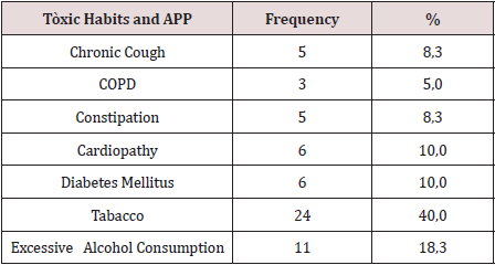
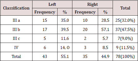
There were operated 78 hernias in 60 patients (18 patients [30.0%] suffered from bilateral hernias, 69 primary hernias and 9 reproduced hernias). The average of age was 55.6 years, the youngest patient was 30 years and the oldest was 77 years, but the majority (12 patients) were in the fifth decade of life. The male sex predominated in 82.9%, which represented a relationship man / woman of 5: 1. 42.9% of patients performed large efforts habitual physicists. The pathological history of the patients (Table 1). It is observed that 24 patients (40.0%) consumed tobacco, and in 11 an excessive consumption of alcohol was collected 18.3%. COPD: Chronic Obstructive Pulmonary Disease (Table 2). The distribution of the series according to the Nyhus classification. Right hernias predominated (55.1%), the indirect variety with large dilation of the ring and destruction of the posterior wall (IIIb) was the most frequent (37 hernias), and 7 femorales hernias and 9 recurrent hernias were operated 73 PET (93.6%) and 5 TAPP (6.4%) were performed. Two of the patients in whom a TEP technique was started were converted to a conventional prosthetic technique by accidental perforation of the peritoneum, passing the CO2 into the peritoneal cavity, and consequently, the loss of the preperitoneal surgical space. Of the 5 TAPP repairs, 3 of them were in the course of a laparoscopic cholecystectomy, and another was the conversion of a failed PET technique. The average surgical time of unilateral hernias was 53.5 min, with a minimum of 25 min and a maximum of 120 min. In bilateral repairs, the average surgical time was 71.3 min, and a minimum of 40 and a maximum was observed. of 110 minutes.

Table 1: Toxicos habits, and personal phalogical history (APP).

lupinepublishers-openaccess-surgery-case-studies-journal

Table 2: Distribution According to the Nyhus Classification.

lupinepublishers-openaccess-surgery-case-studies-journal

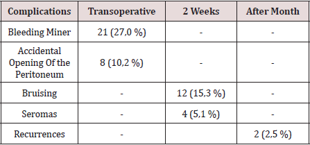
The hospital stay was less than 24 h in 50 patients (71.4%), in 5 it extended from 24 to 48 h, and in 5 to more than 48 hours. The most frequent complication in the transoperative period (Table 3) was minor bleeding in 21 repairs (27.0%) that gave rise to 12 hematomas (15.3%). No complications were observed after the second week, but two patients suffered recurrences (2.5%), more than two months after surgery. The application and evaluation of the VAS scale (Table 4). In the immediate postoperative period, after the patient recovered from anesthesia, 56 individuals (93.3%) were classified as VAS I, and 4 as VAS II. Twenty-four hours after surgery, 14 patients (23.3%) were classified as VAS I, 40 (66.6%) as VASII, 4 patients as VAS III and 2 VAS IV. In the consultation of the first week of postoperatively, 42 patients (70.0%) were classified as VAS I and [10] as VAS II, and two patients with moderate pain (VAS V) appeared in this period. Fifteen days after surgery, 56 individuals (93.3%) were VAS I and a month were 58 (96.6%). The incorporation to the usual activities, including work, was 3 patients a week after surgery, at 15 days they were 19 patients (31.6%) and 54 patients a month (90.0%).

Table 3: Complications.

lupinepublishers-openaccess-surgery-case-studies-journal

Table 4: Evaluation of the Visual Analogy Scale (VAS).

lupinepublishers-openaccess-surgery-case-studies-journal

Discussion

Currently, with the improvement of laparoscopic techniques, these are outlined as safe, reproducible and as a therapeutic option regardless of the age of the patient; nevertheless, the evaluation of the individual must be correct and meticulous in the preoperative period, specifically cardiorespiratory function, since with the TEP method a working space is created between the sheets of the crosssection sheet, richly vascularized, so that absorption and elimination of CO2 is greater than that produced in the peritoneal cavity during the performance of the pneumoperitoneum [10]. Although men predominated, there was a slight increase in women in the series with respect to other authors, [3-5] which could have been due to the inclusion in the study of 3 women who underwent the diagnosis of inguinal hernia, in the course of a laparoscopic cholecystectomy. In laparoscopic practice, it is not uncommon finding of hernia defects diagnosed in the transoperative, in men and women, the latter basically with a history of gynecological disorders. Although the usefulness of hernia repairs in asymptomatic patients is questioned in some articles, [11,12] the authors consider that it would be beneficial for the patient, if conditions permit, the repair of the hernia defect by the TAPP method. The relationship between the hernia disease and the physical efforts, is classic from the Cooper era [1]. In the series, 68% of the patients performed physical activities involving large and medium efforts, and also analyzing the multifactorial character in the pathogenesis of hernia disease, was striking, that approximately half of the patients operated on were smokers, a factor that influences the metabolism of collagen, significantly linked to hernia recurrences [13]. The majority of the repairs were by means of the PET technique, and we consider, as other authors [10,14 -19], that although the TAPP technique brings us closer to the area from a family perspective to the surgeon (peritoneal cavity) and facilitates the so-called “learning curve”, the hernial disease - considering it a parietal defect- should be given solution from this same plane to avoid the likelihood of serious complications of intrabdominal , and to leave the transperitoneal method as a tactical resource when the totally extraperitoneal method is unsuccessful.

The average surgical time was similar to other series [3-6]. It is known that this tends to decrease when the surgical team gains experience [16]. The largest surgical time recorded was in a patient, who was started with a PET technique, but Due to technical difficulties, it was converted to a conventional posterior repair. The fundamental complications were in relation to minor bleeding in the transoperative period and postoperative hematomas. In 3 patients it was necessary to drain the hematoma due to the discomfort caused, however, in the rest of the patients with hematomas and seromas they were treated with conservative measures. In two patients, the recurrence occurred 2 months after surgery, which was interpreted as a technical error. Our results coincide with numerous studies [3-7], that indicate the least postoperative pain of the minimum access techniques, as well as a prompt social and labor reincorporation of the patients. Despite the fact that 70% and 93.3% a week and 15 days postoperatively, respectively, had no pain or discomfort were minimal, only [18] individuals (30%) were incorporated into their usual activities before 15 days. These results contrast with other studies that report a return to work and social activities between 10-15 postoperative days, 4-17- although it is likely that some sociocultural factors are influencing these results. It can be concluded by noting that laparoscopic inguinal hernioplasty is another therapeutic option, mainly in patients with bilateral and reproduced hernias. In the series there were no major transoperative or postoperative complications, only minor bleeding and bruising were present. Most patients were not afflicted by pain 2 weeks postoperatively, however, return to social and labor activities after 15 days was low [18-20].

Read More Lupine Publishers Surgery and Case Studies Journal Articleshttps://surgery-casestudies-lupine-publishers.blogspot.com/


Gallstone Ileus in the Elderly: Still a Challenge, Report of a Case with Review of the Current Literature

  Abstract Introduction:  Gallstone ileus is described as an intestinal obstruction caused by luminal gallstone impaction. It is a mainly ...