Thursday, February 20, 2020

Acute Liver Failure and Thyrotoxicosis Managed with Liver Transplant and Thyroidectomy- Lupine Publishers

Abstract

Acute liver failure and hyperthyroidism are not typically common, although some cases have been reported. The mechanisms involved and optimal management are not well-defined. This article presents the case of a 32-year-old African American female referred for evaluation of abdominal pain and jaundice, with a past medical history of systemic lupus erythematosus and Grave’s disease. She had thyrotoxicosis after administration of contrast and developed acute liver failure culminating in liver transplant and subsequent total thyroidectomy with a favorable outcome.
Keywords: Grave’s Disease; Autoimmune Hepatitis; Thyrotoxicosis; Acute Liver Failure; Liver Transplant; Thyroidectomy; Contrast Induced
Abbrevations: OLT: Orthotopic liver transplant; SLE: Systemic Lupus Erythematosus; TT: Total thyroidectomy; HD: Hospital day; POD: Post-operative day; ANA: Anti-nuclear antibodies; NaMELD: Sodium model for end-stage liver disease; PTU: Propylthiouracil.

Introduction

The association between liver function abnormalities and thyroid dysfunction is well established dating back to at least 1874 when Habershon presented a case of exophthalmic goiter and jaundice to the London Medical Society [1]. The range of liver dysfunction can go from mild elevation of hepatic enzymes to acute liver failure, which is defined as the presence of hepatic encephalopathy within 8 weeks of initial symptoms in a patient without underlying liver disease. Multiple scales have been devised to predict mortality and determine which patients would benefit from orthotopic liver transplant (OLT), with the presence of hepatic encephalopathy as a key indicator [2]. Here we present a case of a 32-year-old female with systemic lupus erythematosus (SLE) and exacerbation of Grave’s disease after iodinated contrast that presented with acute liver failure and thyrotoxicosis requiring OLT and total thyroidectomy (TT) after medical management of both conditions.

Case

A 32-year-old African American female was referred to the emergency room for evaluation of a 3-week history of worsening abdominal pain and jaundice. She also reported loss of appetite and occasional epigastric discomfort. Her past medical history included SLE and Grave’s disease [3]. Both conditions were well controlled, and her medications included multivitamins, hydroxychloroquine and ferrous sulfate. Three weeks prior to admission she underwent a CT scan with iodinated contrast for abdominal pain at an outside hospital. At that time, she received prednisone (60mg PO qd) and diphenhydramine for a presumed diagnosis of autoimmune hepatitis with positive antinuclear antibodies (ANA) at 1:320. Ferritin, alpha 1 antitrypsin, acetaminophen and viral serologies were all negative prior to admission. Her initial vital signs showed BP 110/70 mmHg, HR 134 bpm, RR 20 bpm, T 37°C and SpO2 100%. Physical examination was significant for generalized jaundice and moderate abdominal pain on palpation.
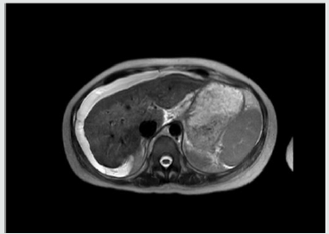
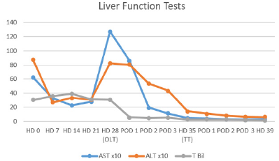
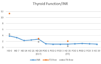
At that time her laboratories were INR 3.8, WBC 36.6 k/uL, K 2.7 mmol/L, lactate 2.4 mmol/L, Alk Phos 187 IU/L, AST 625 IU/L, 872 IU/L, Built 30.2 mg/dL, Bild 21.9 mg/dL, T3 11.3 pg/mL, T4 4.46 ng/dL, TSH 0.02 uIU/mL and ceruloplasmin 26 mg/dL. On HD #11 her 24hr copper excretion was 241.6 ug/d. Her admission Named was 35 and 31 at time of transplant. Renal function remained normal throughout her hospitalization. Trends for her hospital stay are shown in (Tables 1 & 2) After initial stabilization, Doppler ultrasound revealed patent vasculature, and MRI showed nodular appearance of the liver consistent with edema or early cirrhosis (Figure 1). The liver transplant service was consulted and a standard workup for potential liver transplant was started.
Table 1: Liver Function Tests. OLT: Orthotopic Liver Transplant. TT: Total Thyroidectomy.
Lupinepublishers-openaccess-Surgery-Casestudies
Table 2: TSH remained undetectable at <0.02.
Lupinepublishers-openaccess-Surgery-Casestudies
Figure 1: MRI of abdomen showing nodular appearance of liver consistent with edema or early cirrhosis.
Lupinepublishers-openaccess-Surgery-Casestudies
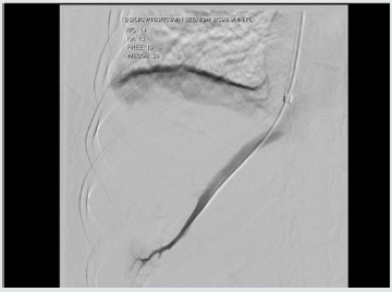
A transvenous liver biopsy on HD #6 was positive for marked active hepatitis with bridging necrosis, parenchymal collapse and minimal focal staining for copper. At the time of biopsy, a hemodynamic study was performed consistent with portal hypertension (Figure 2). Blood cultures were positive for E. coli and urine cultures for E. faecalis on admission, and she received ceftriaxone and meropenem with negative conversion of cultures. Her condition continued to deteriorate with worsening hepatic encephalopathy despite optimal treatment with rifaximin and lactulose. Oral prednisone was continued at 40 mg PO/qd. Her thyrotoxicosis was managed with propranolol and potassium iodide oral solution. An echocardiogram ruled out congestive heart failure. At HD #26 she was admitted to the intensive care unit and due to her condition, it was deemed she was at risk of death within one week and was listed as emergency status 1A on the united network for organ sharing.
Figure 2: Transvenous hemodynamic study consistent with portal hypertension. Free hepatic pressure 19 mmHg, wedge hepatic pressure 26 mmHg, hepatic venous gradient 7 mmHg.
Lupinepublishers-openaccess-Surgery-Casestudies
A suitable donor became available on HD #28 and she underwent OLT with methylprednisolone and mycophenolate induction without complications. She received 250 mcg of potassium iodide the night prior to transplant. Liver explant revealed extensive bridging necrosis, parenchymal collapse, no definite areas of fibrosis and minimal copper staining. She recovered without incidents from transplant and underwent total thyroidectomy on POD #7, she was then started on levothyroxine. She was discharged on HD #39 to an acute rehabilitation facility. At 1-year follow up, she has excellent graft function, remains euthyroid and has not experienced flare ups of SLE.

Discussion

Exposure to iodinated contrast is well known to cause thyroid dysfunction, with an iodine content of 320 to 370 mg/mL it is well above the recommended daily allowance and can precipitate thyrotoxicosis via the Jöb-Basedow phenomenon in patients with Grave’s disease [3]. Hyperthyroidism, particularly in Grave’s disease, is associated with primary biliary cirrhosis and autoimmune hepatitis. In patients without heart failure and hyperthyroidism, the liver biopsy has demonstrated fatty infiltration, cytoplasmic vacuolization, nuclear irregularity and hyperchromatism [4]. Although the exact mechanism for the observed liver-thyroid interactions is unknown, the following have been proposed:
a. systemic effects of thyroid excess,
b. direct toxic effects of thyroid hormone,
c. intrinsic liver and thyroid autoimmune mechanisms,
d. abnormal thyroid metabolism due to liver disease, and
e. subclinical physiologic effects of thyroid hormone [5].
feasible explanation is the presence of a hypermetabolic state with increased hepatic oxygen consumption, but without increases in hepatic blood flow, affecting the centrilobular zones and interfering with bile transport. These findings are consistent with the picture of cholestasis usually present in such cases [6]. In a series of 84 patients with acute liver failure, Anastasiou and colleagues reported a 50% incidence of thyroid hormone abnormalities, with a worse outcome in patients with hyperthyroidism, believed to be secondary to an increase in oxygen consumption and decreased organ perfusion [7]. In the current case, the presence of drug-induced liver injury had to be ruled out, hydroxychloroquine has been associated with acute liver failure requiring liver transplantation, but it appears within two weeks of starting therapy [8]. There is a well-known correlation between anti-thyroid medications and hepatic dysfunction, for example, propylthiouracil (PTU) and methimazole are associated with an incidence of severe liver injury in 0.1% and 0.1-0.2% of patients, respectively [9].
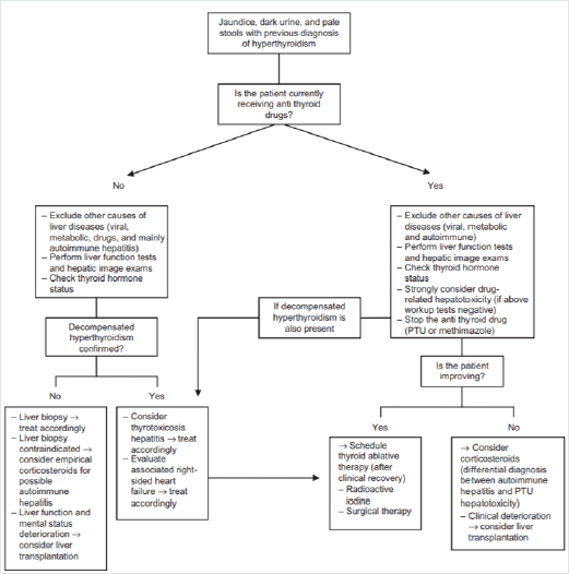
Figure 3: Proposed algorithm for management of patients with symptoms of liver dysfunction and previous diagnosis of hyperthyroidism. [Figure note: Figure obtained from de Campos Mazo, 2013; © 2013 Mazo et al, publisher and licensee Dove Medical Press Ltd. This is an Open Access article which permits unrestricted noncommercial use, provided the original work is properly cited.] [10].
Lupinepublishers-openaccess-Surgery-Casestudies
To our knowledge, our patient was not exposed to these medications. In the absence of established guidelines, a useful algorithm for the management of liver dysfunction with hyperthyroidism has been proposed, (Figure 3) This algorithm is based on a case series of 8 patients in which two cases developed acute liver failure suitable for OLT [10]. Nonetheless, the timing and sequence of OLT and thyroidectomy is not well established. Although it has been proposed not to delay the thyroidectomy as it might represent a potentially life-saving procedure [11]. The incidence of acute liver failure in the setting of hyperthyroidism remains a rare event, with patients undergoing OLT and thyroidectomy even more unusual. In this case report, adequate control of the thyrotoxicosis was achieved prior to transplant with oral potassium iodide and propranolol. In this patient with rapidly progressive liver failure, it appeared clinically prudent to undergo emergent OLT followed by TT.

Conclusion

The presence of ANA in a patient with SLE and Grave’s disease suggest the diagnosis of autoimmune hepatitis, however in this case report, the administration of iodinated contrast and the presence of abnormal copper metabolism made the diagnosis more difficult. The optimal management of these patients is not well established, with only a few cases reported in the literature. Based on our experience, medical stabilization followed by OLT and TT appears to have been an adequate course of treatment. Further studies are needed to help determine guidelines./p>


To know more about Open access publishers click on Lupine Publishers


Friday, February 7, 2020

Quantification of MicroRNAs for the Diagnostic Screening of Colon Cancer in Human Stool by Absolute Digital(d)PCR*| Lupine Publishers


Abstract


There is currently no validated micro(mi)RNA diagnostic stool test to screen for colon cancer (CC) on the market because of the complexity of fecal density, vulnerability of stool to daily changes, and the presence of three sources of miRNAs in stool (cell-free from fecal homogenates, exsosomal miRNAs from fecal exosomes, and fecal colonocytes). By employing earlier on a microarray miRNA experiment, using Affymetrix GeneChip miRNA 2.0 Arrays, on immunocaptured and enriched stool colonocytes of 15 subjects [three healthy controls and twelve colon cancer patients [three TNM stage 0-1 (e.g., polyps  1 cm, villous or tubvillous, or with high grade dysplasia), three stage 2, three stage 3, and three stage 4] in triplicates, this allowed for selection of a smaller panel of 14 preferentially expressed mature miRNAs associated with colon cancer (12 Up-Regulated, miR-19a, miR-20a, miR-21, miR-31, miR-34a, miR-96, miR-106a, miR-133a, miR-135b, miR-206, miR-224 and miR-302; and 2 Down-Regulated, miR-143 and miR-145). Then carrying out an absolute quantitative digital PCR on these 15 stool samples from TNM stages 0-4 on total small RNA extracted by immunocapture, followed by RT that employed a Custom TaqMan® miRNA Reverse Transcription (RT) Kit and TaqMan RT Primer Pool, and absolute quantification of miRNAs, in copies/μl, measured using a chip-based Absolute QuantStudio 3D Digital PCR analysis, allowed for validating the microarray results. To ensure that human and not bacterial small total RNA was chosen, coextraction protocols with E. coli K1 strain RS18 was carried out, followed by comparing Agilent electrophoretic patterns with human and bacterial electrophoretic patterns, and also random samples were sequenced using mRNA/miRNA sequencing, to ensure that human and not bacterial mRNA was chosen.

Introduction


Quantitative dPCR miRNA data presented in herein, show that the quantitative changes in the expression of a few mature miRNA genes in stool, which are associated with right and left colon cancer, would provide for a more convenient, sensitive and specific diagnostic screening molecular markers, more useful than markers currently available on the market, such as the low-sensitivity (<15%) fecal occult blood test(FOBT); result in better compliance; and is more economical than the invasive and expensive colon cancer colonoscopy exam, resulting in a higher probability of curing that cancer, if detected at the early TNM stages, and which becomes incurable and deadly if not diagnosed before metastasis.

Advantages of Using a MiRNA Diagnostic Colon Cancer Screening Test

The expression of individual genes may be altered by mutations in the DNA, or by a change in their regulation at the RNA or protein levels [1]. Epigenetic silencing is an important mechanism that contributes to gene inactivation in colorectal cancer (CRC) [2]. Analysis of promoter methylation of hypermethylated in cancer 1 (HIC1) gene in human stool showed it to be highly specific (98%) for both colon adenoma and carcinoma [3], but the sensitivity was quite low (31% for adenoma & 42% for all cancer), suggesting that an epigenetic marker only is not adequate for an accurate diagnostic screening, but a combination of genetic and epigenetic markers would be required to reliably identify CRC at an early disease stage [4]. Working with the stable DNA has been relatively easy compared to working with the fragile RNA molecule [1].A study by scientists at Exact Sciences Corp, Marlborough, MA, which markets a mutation-based DNA test “Cologuard”, assessed a newer version of a fecal DNA test for CRC screening using a vimentin methylation marker and another mutation DY marker plus nondegraded DNA in a limited sample of 44 CRC patients and 122 normal controls [5].
It cited a sensitivity of 88% and a specificity of 82%, only for advanced cancer, but not for the early adenoma stage. Besides, DNA mutation tests are not cost-effective, as screening for multiple mutations is expensive because these demanding mutation tests are not automated and are labor intensive. In addition, mutation detection in oncogenes and suppressor genes suffers from: a) the detection of mutations in these genes in fewer than half of large adenomas and carcinomas, b) the detection of gene mutations in non-neoplastic tissues, c) mutations found only in a portion of the tumor, and d) mutations often produce changes in the expression of many other genes [6,7]. Protein-based methods are currently not suited for screening and early diagnosis, either because proteins are not specific to one tumor or tissue type (e.g., CEA), their susceptibility to proteases, current lack of means to amplify proteins, no function is known for more than 75% of predicted proteins of multicellular organisms, there is not always a direct correlation between protein abundance and activity, and most importantly because detection of these markers exfoliately often signifies the presence of an advanced tumor stage. The dynamic range of protein expression in minimally-invasive body fluids (e.g., blood) is as large as 1010. Moreover, mRNA levels do not necessarily correlate with protein expressions.
Protein microarray studies revealed that protein expression vastly exceeds RNA levels, and only post translationally modified proteins are involved in signal transduction pathways leading to tumorigenesis. There is no well-documented protein test that has been shown in clinical trials to be a sensitive and a specific indicator of colon neoplasia, especially in early stages [8]. A serum proteomic study employing liquid chromatography (LC)-mass spectrometry (MS) carried out in a non-biased fashion failed to differentiate between individuals with large adenoma ( 1 cm) and normal individuals [9]. Compared to nucleic acids, proteomic research is a newer discipline; therefore, it will take considerable time to identify and validate proteins suitable for use as clinical markers, and resolve issues of bias and validations [10].On the other hand, a transcriptomic mRNA approach, has been shown to detect both adenomas and colon carcinomas with high sensitivity and specificity in preliminary studies [1], but no randomized, standardized, blinded prospective clinical studies have been carried out to validate the superiority of the mRNA approach.
A study indicated that a combination of a transcriptomic mRNA and miRNA expression signatures improves biomolecular classification of CRC [11]. Furthermore, not only does miRNAs regulate mRNA, but they also regulate protein expression. Two studies have shown that a single miRNA act as a rheostat to fine tune the expression of hundreds of proteins [12,13]. Hence, for CRC screening, miRNA markers are much more comprehensive and preferable to a DNA-, epigenetic-, mRNA- or a protein-based marker [14-18]. An added advantage for the use of the stable, nondegradable miRNAs by PCR expression, by chip-based methods, is its being automatable, making them much more economical and more easily acceptable by laboratory personnel performing these assays [4]. The discovery of small non-coding protein sequences,17-27 nucleotides long RNAs (microRNAs), has opened new opportunities for developing a non-invasive screening test for early diagnosis of many cancers. The latest miRbase release 22 on, March 12, 2018 [http://ww.mirbase.org] indicates the total number of miRNAs labeled “high confidence” has increased by 168, to 1996, than in the previous release [19].
MiRNA functions seem regulate development [20], apoptosis [21], and specific miRNAs are essential in oncogenesis [22,23], effective in classifying solid [24-26] and liquid tumors [27,28], and could serve as oncogenes or suppressor genes [29]. MiRNA genes are frequently found at fragile sites, as well as minimal regions of loss of heterozygosity, or amplification of common break-point regions [30], implying their involvement in carcinogenesis. MiRNAs have potential to serve as biomarkers for cancer diagnosis, prognosis and/or response to therapy [31,32]. Profiles of miRNA expression differ between normal and tumor tissues (33,34), suggesting that their expression profiles cluster similar tumor types together more accurately than expression profiles of protein-coding mRNA genes [33,34]. A study that examined global expression of 735 miRNAs in 315 samples of normal colonic mucosa, tubulovillus adenomas, adenocarcinomas proficient in DNA mismatch repair (pMMR), and defective in DNA mismatch repair (dMMR) representing sporadic and inherited CRC stages I-IV suggest involvement of common biologic pathways in pMMR and dMMR tumors in spite of the presence of numerous molecular differences between them, including differences at the miRNA level; indicating the need to pay attention to mismatch DNA repair (MMR) [34] .
Unlike screening for large numbers of messenger (m)RNA [1], a modest number of miRNAs is used to differentiate cancer from normal [35], and unlike mRNA, miRNAs in stool remain largely intact and stable for detection [36], therefore, leading to conclude that miRNA molecules are better markers to use for developing a reliable noninvasive diagnostic marker screen for colon cancer [14-18], since: a) the presence of the bacterium Escherichia coli does not hinder detection of miRNA by a sensitive technique such as dPCR [36], and b) the miRNA expression patterns are the same in primary tumor, or diseased tissue, as in stool samples [35-37]. The gold standard to which the miRNA test is compared to, has been “colonoscopy”, obtained from patients’ medical records [38]. However, because the low sensitivity guaiac FOBT is still the most commonly used screen in annual checkups (www.cancer.org) [39], this test should also be included for comparison with the proposed dPCR diagnostic miRNA screening approach in human stool.

Advantages of Stool Over Other Testing Media

Stool testing has several advantages over other colon cancer screening methods a s it is truly noninvasive and requires no unpleasant cathartic preparation, formal health care visits, or time away from work or routine activities [40-43]. Unlike sigmoidoscopy, it reflects the full length of the colorectum and samples can be taken in a way that represents the right and left side of the colon. It is also believed that colonocytes are released continuously and abundantly into the fecal stream, contrary to blood that is released intermittently as in guaiac fecal occult blood test (FOBT) [39]; therefore, this natural enrichment phenomenon partially obviates the need to use a laboratory-enrichment technique to enrich for tumorigenic colonocytes, as for example when blood is used for screen testing [44]. Furthermore, because testing can be performed on mail-in-specimens, geographic access stool screening is essentially unimpeded [4]. The American Cancer Society (ACS) has recognized stool-based molecular testing as a promising screening technology for CRC (www.cancer.org).
Isolation of colonocytes from stool, and comparing the Agilent electrophoretic (18S and 28S) patterns to those obtained from total RNA extracted from whole stool [45-47], and differential lysis of colonocytes by RT lysis buffer (Qiagen), could be construed as a validation that the electrophoretic pattern observed in stool (18S and 28S) is truly due to the presence of human colonocytes, and not due to stool contamination with Escherichia coli (16S and 23S). Taking into account that some exsosomal RNA will be released from purified colonocytes into stool, attempts must be made to correct for exsosomal RNA effect [48].

MiRNA dPCR Study Design

To test miRNAs as reliable, quantitative, sensitive and specific diagnostic biomarkers, for early non-invasive screening of colon cancer, using absolute dPCR test, preliminary work must be validated in a study using a nested case control epidemiology design and employing a prospective specimen collection, retrospective blind evaluation (Probe) of control subjects and test colon cancer patients, as specifically delineated by the National Cancer Institute’s Early Detection Research Network http://edrn.nci.nih.gov for cancer biomarker discovery studies. Selection of 14 miRNAs, 12 of them showed increased expression and 2 showed decreased expression for analysis of absolute miRNAs expression by a chipbased digital (d) PCR test is presented in Tables 1 & 2, and Figure 1. Absolute Quantitative Digital PCR Approach Digital PCR is a new approach to miRNAs quantification that offers an alternate method to qPCR for absolute quantification, by partitioning a sample of DNA or cDNA into many individual, parallel PCR reactions; some of these reactions contain the target molecule (positive), while others do not (negative).
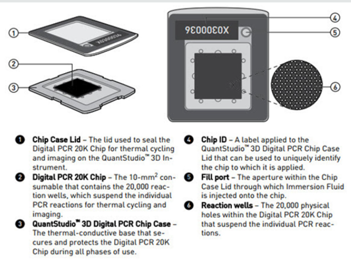
Figure 1: Diagram illustrating QuantStudioTM 3D Digital PCR System Chip; ChipCase Lid (1); Digital PCR 20K 10 mm2 nanofluidic v2 chip (2), which contains 20,000 reaction wells; QuantStudioTM 3D Digital PCR Chip Case (3); Chip ID (4); Fill port (5); and Reaction wells, the 20,000 physical holes that suspend individual PCR reactions.
Lupinepublishers-openaccess-Surgery-Casestudies
Table 1: Characteristics of Fourteen Up- or Down-Regulated MicroRNAs in Human Stool.
Lupinepublishers-openaccess-Surgery-Casestudies
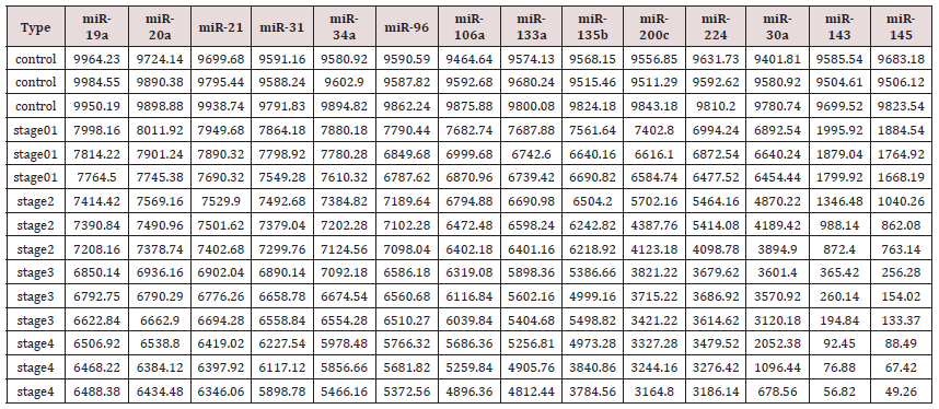
Table 2: Absolute Quantification of Up-/Down- Regulated miRNAs in Stool by QuantStudioTM 3D Chip-Based Digital PCR.
Lupinepublishers-openaccess-Surgery-Casestudies
A single molecule can be amplified a million-fold or more. During amplification, TaqMan chemistry with dye-labeled probes is used to detect sequence-specific targets. When no target sequence is present, no signal accumulates. Following PCR analysis, the fraction of negative reactions is used to generate an absolute count of the number of target molecules in the sample, without the need for standards or endogenous controls. In conventional qPCR, the signal from wild-type sequences dominates and obscures the signal from rare sequences [49-51]. By minimizing the effect of competition between targets, dPCR overcomes the difficulties inherent to amplifying rare sequences and allows for sensitive & precise absolute quantification of the selected miRNAs. Applied Biosystem QuantStudio™ 3D instrument used in this research study, only performs the imaging and primary analysis of the digital chips. The chips themselves must be cycled offline on a Dual Flat Block GeneAmp® 9700 PCR System. or the ProFlex™ 2x Flat PCR System. The QuantStudio™ 3D Digital PCR System can read the digital chip in less than 1 minute, following thermal cycling [45].
It allows for one sample per chip; although, duplexing allows for analsis of two targets per chip. Sample prep for digital PCR is no different than for real-time qPCR, when using the QuantStudio™ 3D Digital PCR System. To figure out the concentration of cDNA stock from results, if one includes all of the necessary dilution factors into the AnalysisSuite™ software, the software will give the copies/μL in the stock. There are 2 dilutions that one needs to take into account:
a) The first is the dilution of the sample in the reaction,. and
b) The second is the dilution of the stock that one makes before adding it to the digital PCR reaction.
For example, if one wants to add 1 μL of a sample that has been diluted 1:10 from the stock. Thus, if one adds 1 μL of his/her sample to a 16 μL (final volume) reaction, the dilution factor of the sample is 1:16 or 1/16 = 0.0625. Since the stock has also been diluted 1:10 (0.1), one also need to factor this in. The final dilution factor to enter into the software is 0.0625 x 0.1 = 0.00625 (1:160). One can use either annotation to indicate the dilution factor in the AnalysisSuite™ software. If one enters that value into the “Dilution” column, the software will give the copies/μL in the starting material (stock).
The Poisson Plus algorithm for projects that contain QuantStudio™ 3D Chips with target, quantities >2000 copies/μL. The Poisson Plus algorithm corrects for well-to-well load volume variation, on a per Chip basis. This becomes important at higher target concentrations. There is also an option to export the Chip data as XML on the Export tab-thousands of discrete subunits prior to amplification by PCR, each ideally containing either zero or one (or at most, a few) template molecules [50]. Each partition behaves as an individual PCR reactions –as with real-time PCR-fluorescent FAM probes [or others, as VIC fluorescence]. Samples containing amplified products are considered positive (1, fluorescent), and those without product –with little or no fluorescence (i.e., are negative, 0). The ratio of positives to negatives in each sample is the basis of amplification. Unlike real-time qPCR, dPCR does not rely on the number of amplification cycles to determine the initial amount of template nucleic acid in each sample, but it relies on Poisson Statistics to determine the absolute template quantity.
The unique sample partitioning step of dPCR, coupled with Poisson Statistics allows for higher precision than both traditional end point PCR. and qPCR methods; thereby allowing for analysis of rare miRNA targets quantitatively and accuratley [47,48]. The use of a nanofluidic chip, shown below, provides a convenient and straight forward mechanism to run thousands of PCR reactions in parallel. Each well is loaded with a mixture of sample, master mix, and Applied Biosystems TaqMan Assay reagents, and individually analyzed to detect the presence (positive) or absence (negative) of an endpoint signal. To account for wells that may have received more than one molecule of the target sequence, a correction factor is applied using the Poisson model. It features a filter set that is optimized for the FAM™, VIC®, and ROX™ dyes, available from Life Technologies [46]. The chips themselves must be cycled offline on a Dual Flat Block GeneAmp® 9700 PCR System. or the ProFlex™ 2x Flat PCR System. The QuantStudio™ 3D Digital PCR System can read the digital chip in less than 1 minute, following thermal cycling [45].
It allows for one sample per chip; although, duplexing allows for analsis of two targets per chip. Sample prep for digital PCR is no different than for real-time PCR, when using the Quant Studio™ 3D Digital PCR System. To figure out the concentration of cDNA stock from results, if one includes all of the necessary dilution factors into the AnalysisSuite™ software, the software will give the copies/sL in the stock. There are 2 dilutions that one needs to take into account:
a) The first is the dilution of the sample in the reaction,. and
b) The second is the dilution of the stock that one makes before adding it to the digital PCR reaction.
For example, if one wants to add 1 μL of a sample that has been diluted 1:10 from the stock. Thus, if one adds 1 μL of his/her sample to a 16 μL (final volume) reaction, the dilution factor of the sample is 1:16 or 1/16 = 0.0625. Since the stock has also been diluted 1:10 (0.1), one also need to factor this in. The final dilution factor to enter into the software is 0.0625 x 0.1 = 0.00625 (1:160). One can use either annotation to indicate the dilution factor in the AnalysisSuite™ software.
If one enters that value into the “Dilution” column, the software will give the copies/μL in the starting material (stock). The Poisson Plus algorithm for projects that contain Quant Studio™ 3D Chips with target, quantities >2000 copies/μL. The Poisson Plus algorithm corrects for well-to-well load volume variation, on a per Chip basis. This becomes important at higher target concentrations. There is also an option to export the Chip data as XML on the Export tab-thousands of discrete subunits prior to amplification by PCR, each ideally containing either zero or one (or at most, a few) template molecules [47]. Each partition behaves as an individual PCR reactions –as with real-time PCR—fluorescent FAM probes [or others, as VIC fluorescence]. Samples containing amplified products are considered positive (1, fluorescent), and those without product –with little or no fluorescence (i.e., are negative, 0). The ratio of positives to negatives in each sample is the basis of amplification. Unlike real-time qPCR, dPCR does not rely on the number of amplification cycles to determine the initial amount of template nucleic acid in each sample, but it relies on Poisson Statistics to determine the absolute template quantity.
The unique sample partitioning step of dPCR, coupled with Poisson Statistics allows for higher precision than both traditional and qPCR methods; thereby allowing for analysis of rare miRNA targets quantitatively and accuratley [47]. The use of a nanofluidic chip, shown below, provides a convenient and straight forward mechanism to run thousands of PCR reactions in parallel. Each well is loaded with a mixture of sample, master mix, and Applied Biosystems TaqMan Assay reagents, and individually analyzed to detect the presence (positive) or absence (negative) of an endpoint signal. To account for wells that may have received more than one molecule of the target sequence, a correction factor is applied using the Poisson model. It features a filter set that is optimized for the FAM™, VIC®, and ROX™ dyes, available from Life Technologies [46]. A workflow of the dPCR procedure by the QuantStudioTM 3D Digital PCR System is presented in (Figure 2) Workflow of a digital miRNAs PCR for colon cancer profiling in human colon tissue or stool samples.
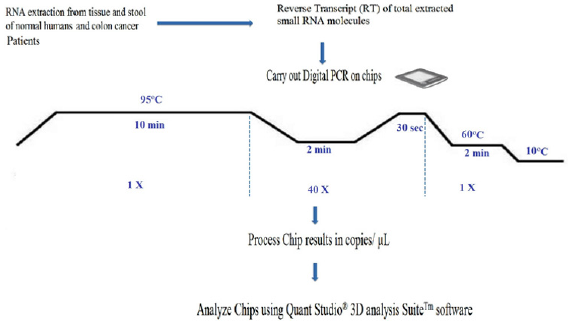
Figure 2: Workflow of a digital miRNAs PCR for colon cancer profiling in human colon tissue or stool samples.
Lupinepublishers-openaccess-Surgery-Casestudies

Digital PCR, however, has Several Tips to Follow:

i. A rough estimate of the concentration of miRNAs of interest has to be first carried out, in order to make appropriate dilutions, so that not too many partitions will get multiple copies that prevent accurate calculation of the copy number of miRNAs of interest;
ii. Non-template controls and a RT negative control must be set up for each miRNA, when using a “primer pool method” for retro-transcription;
iii. A chip-based dPCR method requires less pipetting steps, which reduces potential PCR contamination compared to another type of dPCR marketed by Bio-Rad Laboratories, thus called “Bio-Rad’s droplet digital PCR”, which requires multiple pipette transfers that potentially increase the risk of contamination [47], and
iv. Quant StudioTM 3D chip has 20,000 fixed reaction wells, whereas Bio-Rad’s droplet PCR relies upon the generation of droplets;
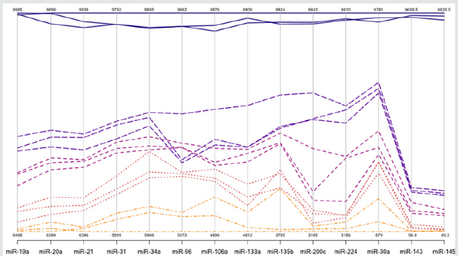
a step that could be extremely variable, as reported by Miotto et al (11,48) Absolute dPCR data tabulated in Tables 1 & 2, and presented graphically in Figure 1 below, which show 14 preferentially expressed mature miRNAs associated with colon cancer (12 Up-Regulated, miR-19a, miR-20a, miR-21, miR-31, miR- 34a, miR-96, miR-106a, miR-133a, miR-135b, miR-206, miR-224 and miR-302; and 2 Down-Regulated, miR-143 and miR-145) in stool samples from healthy controls, and stages 0-1 to 4 individuals with colon cancer . Standard deviations (sd) obtained from the one-way ANOVA, using the 5 level factor Type (normal, stage01, stage2, stage3, stage4) were calculated. The adjusted R-squared values representing the proportion of variation explained by Type are also reported. Type was statistically significant for every gene; all p-values were less than 0.000001 (no adjustments for multiple comparisons). These data are tabulated in Table 3 and shown graphically in Figure 1. For each gene on the graph in Figure 1, the min and max have been shown, in order to make the presentation clearer. At top left is high exxpression Value of 9985, which is the maximum value for that gene, at the bottom one finds the value for the minimum the colors range from dark blue (control) to orange (stage 4).
Table 3: Representation of SDs and R2 for miRNAs tested by absolute digital PCR.

Lupinepublishers-openaccess-Surgery-Casestudies
The groups are also distinguished by line type: control (solid), stage 0-1 (long dash), stage 2 (dash), stage 3 (dot), stage 4 (dash nd dot). The figure is a parallel coordinate plot made in R, using the package MASS. For statistical analysis. Innovation and Clininical Significance of the dPCR-miRNA Diagnostic Stool Screening Approach Innovation lies in the collective use of many methods, such as: immunoparamagnetic beads to capture colonocytes from the harsh, noninvasive stool environment, whose extracted fragile total small RNA is stabilized shortly after stool excretion by commercial kits so it does not ever fragment, followed by standardized analytical quantitative miRNA dPCR-chip profiling in stool samples, which are neither labor intensive, nor require extensive sample preparation, to develop a panel of few stable miRNAs for absolute quantitative diagnostic screening of early sporadic colon cancer (stage 0-1), cheaper, with higher sensitivity and specificity than any other colon cancer screening test on the market . Isolation of colonocytes from stool samples is needed to provide a quantitative estimate of how our proposed miRNA method performs. Although we may miss exosome RNA, a parallel test could also be carried out on miRNAs obtained from stool samples to compare the extent of loss when colonocytes are only used, and an appropriate correction for exsosomal loss can be made [48]. Figure 3 Absolute Quantification of Up- or Down-Regulated miRNAs in Human Stool by Quant Studio TM 3D Digital PCR Chip System.
Figure 3: Absolute Quantification of Up- or Down-Regulated miRNAs in Human Stool by QuantStudioTM 3D Digital PCR Chip System.
Lupinepublishers-openaccess-Surgery-Casestudies

Acknowledgments


We express our thanks to participating volunteers who provided stool samples for the study; Dr. Paul W. Vos of the Department of Biostatistics, East Carolina University for Statistical Analysis, and Dr. Clark D. Jeffries at Renaissance Computing Institute, University of North Carolina at Chapel Hill for his insight on bioinformatics’ analysis.

 Read More Lupine Publishers Surgery Journal Articles : https://surgery-casestudies-lupine-publishers.blogspot.com/
Read More Lupine Publishers Articles : https://lupinepublishers.blogspot.com/

Friday, January 24, 2020

Mini OPCAB Coronary Artery Bypass Surgery Plus Medical Treatment: An Option for High Risk Coronary Patients_Lupine Publishers

Surgery Journal| Lupine Publishers

Abstract


MINI OPCAB surgery (Xiphoid Approach) is a surgical technique in which the left internal mammary artery is bypassed to the Anterior Descending Artery (ADA) by a medial inferior sternotomy Fourteen high risk patients with multiple coronary disease with a preoperative logistic Euroscore of 10.86 were operated and follow up with medical treatment and strictly control of risks factors MACE at 80 months was 0% and Survival at 7 years 82% (KM) Although is an alternative the combination of Mini OPCAB operation plus medical treatment in high risk. Patients with multiple vessels coronary disease, more experience is needed to confirm this initials results. Statistical analysis applied the student test (SPSS program), with p<0.05 were considered significant
Keywords: Mini Opcab; Minimally Invasive Coronary Surgery; Coronary High Risk Patients

Introduction


In high-risk patients with multiple vessel disease who are not candidates for conventional surgery with extracorporeal circulation or for percutaneous procedures as a single treatment, the alternative of left mammary artery to left internal descending artery bypass graft surgery without extracorporeal circulation offers advantages over medical treatment [1,2].
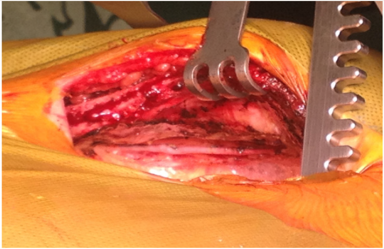
Figure 1:
Lupinepublishers-openaccess-Surgery-Casestudies
Figure 2:
Lupinepublishers-openaccess-Surgery-Casestudies
a) The MIDCAB operation
b) Is effective to treat high risk patients with multiple vessel disease. Greater long-term follow-up is necessary to clarify the indications and validate the procedure for this type of patients.
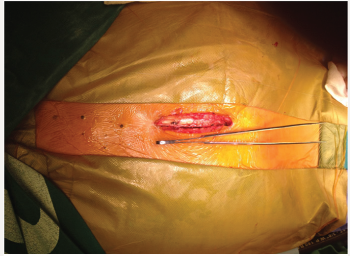
c) MINI OPCAB surgery (Xiphoid Approach) is a surgical technique in which the left internal mammary artery is bypassed to the Anterior Descending Artery (ADA) by a medial inferior sternotomy approach in the 3rd or 4th intercostal space, leaving intact the sternal manubrium (Figures 1 & 2). Long-term results have already been published, reaching 82% survival at 12 years (Kaplan-Meier).
d) This presentation describes the experience with this surgical technique in our institution [3,4].

Materials and Methods


Fourteen high risk patients with multiple vessel coronary artery disease with mean age of 71.07 years (±9.051, 95% CI), 21% women and mean preoperative Logistic EuroSCORE of 10.68 (±5.407, 95% CI), were operated-on in the last 7 years, followed up in our institution with strict medical treatment and control of risk factors.

Result


Operative mortality in this series was 0%, the incidence of perioperative infarction was 0%, the average duration of surgery was 2 hours and 20 minutes; 10 (71%) patients were extubated in the operating room and average hospital stay was 2 days and 11 hours. Following the intervention, one patient received a stent in the right coronary artery and another in the circumflex artery for presenting large arteries with severe lesions. In this group of patients, major adverse cardiovascular events were 0% at 80 months. Survival rate was 82% at 7 years (Kaplan-Meier); an 85-year-old woman died 5 years after surgery due to stroke.

Conclusion


The combination of a MINI-OPCAB surgery for bypass of the left internal mammary artery to the left anterior descending artery [5,6] together with an adequate medical treatment and a hybrid treatment when the right coronary artery or circumflex artery are of high caliber, is a viable option for elderly and high-risk patients. More experience is needed to confirm these initial results.


Abstract


MINI OPCAB surgery (Xiphoid Approach) is a surgical technique in which the left internal mammary artery is bypassed to the Anterior Descending Artery (ADA) by a medial inferior sternotomy Fourteen high risk patients with multiple coronary disease with a preoperative logistic Euroscore of 10.86 were operated and follow up with medical treatment and strictly control of risks factors MACE at 80 months was 0% and Survival at 7 years 82% (KM) Although is an alternative the combination of Mini OPCAB operation plus medical treatment in high risk. Patients with multiple vessels coronary disease, more experience is needed to confirm this initials results. Statistical analysis applied the student test (SPSS program), with p<0.05 were considered significant
Keywords: Mini Opcab; Minimally Invasive Coronary Surgery; Coronary High Risk Patients

Introduction


In high-risk patients with multiple vessel disease who are not candidates for conventional surgery with extracorporeal circulation or for percutaneous procedures as a single treatment, the alternative of left mammary artery to left internal descending artery bypass graft surgery without extracorporeal circulation offers advantages over medical treatment [1,2].
Figure 1:
Lupinepublishers-openaccess-Surgery-Casestudies
Figure 2:
Lupinepublishers-openaccess-Surgery-Casestudies
a) The MIDCAB operation
b) Is effective to treat high risk patients with multiple vessel disease. Greater long-term follow-up is necessary to clarify the indications and validate the procedure for this type of patients.
c) MINI OPCAB surgery (Xiphoid Approach) is a surgical technique in which the left internal mammary artery is bypassed to the Anterior Descending Artery (ADA) by a medial inferior sternotomy approach in the 3rd or 4th intercostal space, leaving intact the sternal manubrium (Figures 1 & 2). Long-term results have already been published, reaching 82% survival at 12 years (Kaplan-Meier).
d) This presentation describes the experience with this surgical technique in our institution [3,4].

Materials and Methods


Fourteen high risk patients with multiple vessel coronary artery disease with mean age of 71.07 years (±9.051, 95% CI), 21% women and mean preoperative Logistic EuroSCORE of 10.68 (±5.407, 95% CI), were operated-on in the last 7 years, followed up in our institution with strict medical treatment and control of risk factors.

Result


Operative mortality in this series was 0%, the incidence of perioperative infarction was 0%, the average duration of surgery was 2 hours and 20 minutes; 10 (71%) patients were extubated in the operating room and average hospital stay was 2 days and 11 hours. Following the intervention, one patient received a stent in the right coronary artery and another in the circumflex artery for presenting large arteries with severe lesions. In this group of patients, major adverse cardiovascular events were 0% at 80 months. Survival rate was 82% at 7 years (Kaplan-Meier); an 85-year-old woman died 5 years after surgery due to stroke.

Conclusion


The combination of a MINI-OPCAB surgery for bypass of the left internal mammary artery to the left anterior descending artery [5,6] together with an adequate medical treatment and a hybrid treatment when the right coronary artery or circumflex artery are of high caliber, is a viable option for elderly and high-risk patients. More experience is needed to confirm these initial results.

Read More Lupine Publishers Surgery Journal : https://surgery-casestudies-lupine-publishers.blogspot.com/

Read More Lupine Publishers Articles : https://lupinepublishers.blogspot.com/
Wednesday, January 22, 2020

Lupine Publishers |Scientific Student Societies: A Way of Scientific Research Vocations Boosting

Lupine Publishers|  Journal of Surgery


Abstract

Born in Russia at the end of the XIXth century the Scientific Student Societies have developed a rather original way of initiation and involvement of the young in the scientific research during High School and University cursus. All through the XXth century and up to now, they have given several generations of Russian scientists an adequate pre formation consisting not only of skill acquisition in their future speciality but also in planning, realization and organization of scientific research and sharing of its results. Their popularity was also due to the high degree of initiative and autonomy left to the students.

Introduction

In our days, when most of young people with a diploma of physician are no more inspired by a scientific career possibility, and though Occidental World has general negative opinion about Russia and its institutions, it is not forbidden to exhume some moments which have proved efficacy and may be useful for.com now. Such an interesting institution (from my point of view) was the Scientific Student Societies (SSS) [1], existing in all the High Schools and Universities, but may be especially activated in Medicine Faculties. They were created in the end of XIXth century by famous scientists such as VI Vernadski (Figure1) and his colleagues in Sankt Petersburg (1882) and in the beginning of the XXth century by NE Jukovski (Figure 2) in Moscow (1909) [1-4]. In the 30-ies they were reactivated and widespread through all the country. After the 2nd World War, SSS were also working in the other countries of the “East Bloc”. The links and experience exchanges were current between them (International meetings and conferences, scientific information share, student exchanges and so on). Presently in the XXIth century they have not disappeared: “NIRS” - student scientific research work - remains a preoccupation of the academic staff in Universities and High Schools as well as different Ministries of the Russian Federation. Student scientific associations still exist, may be a little modified, sometimes independently as “Meridian”, “Luch” (that means ray or beam). Books including scientific articles written by students are still published [5-7], regional and national competitions are organized.
Figure 1: Vladimir Vernadski (1863-1945) famous chemist and mineralogist, creator of the first student scientific circles in Russia.
Lupinepublishers-openaccess-Surgery-Casestudies
Figure 2: Monument to NE Jukovski (1847-1921) - The “Father of Russian Aviation” - in the Entry Square of the Petrovski Palace (Aviation school) in Moscow.
Lupinepublishers-openaccess-Surgery-Casestudies

Description and Commentaries

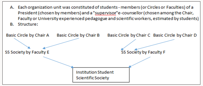
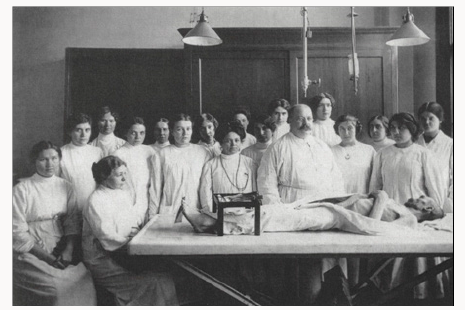
The SSS aims were: to popularize scientific research among students, to find out the most talented among them and to help them to develop towards an eventual scientific career. It was also a mean for students to acquire deeper knowledge in one or several disciplines and to choose their future speciality. The Student Scientific Circles eventually provided the departments with qualified helpers, as a just return for the teaching staff mobilisation during the young researcher’s formation. The structure schema of the SSS is presented in Figure 3. At the basis level was the Circle. As soon as they wanted, students were invited to join a student scientific circle by the department of their choice and to begin their initiation according to their trends with one of the assistant or docent. At this level, the students learned step by step how to manage scientific literature, to review it, to deepen their knowledge’s in the chosen subject, to acquire technical skills and, last but not least, to participate to the research of their mentor/supervisor, or even to start their own investigations on their personal research topic.*NB. The work performed within the SSS activity differed from end of cycle obligatory works, but the members of SSS were allowed to include their personal data in the end of cycle work, if it was adequate. The students - members of a circle - elected their president - responsible who organized periodic reunions for reviewing the work performed by the circle members. During these meetings experienced assistants and docents or professors were invited to present lectures or demonstrations helping the students to assess the peculiarities and the research specifics of the discipline (Figure 4). The best student ‘works were selected and recommended for reporting at Faculty and inter Faculties student scientific conferences.
Figure 3: Schema of a Student Scientific Society structure.
Lupinepublishers-openaccess-Surgery-Casestudies
Figure 4: Professor SHOR GV (1812-1944) - well known Pathologist - with students at a meeting of the student scientific circle of Pathologic Anatomy in the 30-ties.
Lupinepublishers-openaccess-Surgery-Casestudies
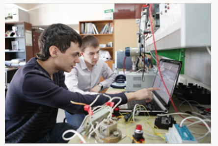
The next level was Faculty and University or High School Scientific Student Society. The department circles joined forming faculty and institute Student Scientific Societies (SSS), which were gathered as University or High School SSS. They were handed by students and only supervised by a responsible chosen among docents and professors of the Faculty. Students elected their president and delegates for contacts with other faculties, institutes of the country and of foreign countries. They organized faculty conferences, Inter-faculty scientific sessions. Their activity was financed either by the Professional Union of their School or by the University budget. The most promising students - members of SSS were further recommended for scientific work either in the department where they started their work, or in Scientific Research Institutes and other High School Faculties (according to the young specialist’s will and the possibilities of the aforementioned institutions). The Ministries took into account the SSS activity of the young specialist when his future was considered after the end of his cursus. Besides career start consideration, other awards were current: scientific books, scientific travels, being the co-author of a “chief’s paper, and so on. So the students made acquaintance not only with the individual searching process itself (Figure 5), but also with elements of organizing scientific research, co-operative investigation, discussion and peer evaluation of its results both by other students and by the Faculty staff scientists. Students also learned to communicate at different levels and, if they wanted, to prepare themselves to a scientific career. If not, nevertheless the aptitudes obtained during their work in the SSS was precious for their further professional activity: acquaintance with special literature research methods, reviewing and critics of scientific publications, acquisition of some technical skills, capacity to plan scientific research and organize scientific meetings.
Figure 5: Students at Research work in a Technical High School in the 80-ies.
Lupinepublishers-openaccess-Surgery-Casestudies
It is estimated that about 20% of the students attended the SSS. Among them about 30% have presented a valuable work (compilation, fundamental or applied research). Most of the exmembers of SSS have pursued a career in the previously chosen specialty. In Russia, all through the years 1930-1990 and up to now most of those who have followed a successful scientific or pedagogic career, were ancient members of the SSS. (That does not mean that without SSS a scientific career was not possible). Many of the students, even foreigner hosts, who have passed through this “school” have been later eminent scientists of their countries (for instance: Academician VI Shumakov - the first Director of the Transplantation Institute in Moscow, Professor VM Filipov - present Rector of the Russian University od People Friendship; Professor R Roman Ramos - Dean of the Medicine Faculty of the Mexico Autonomous University)

Conclusion

The SSS have given several generations of motivated Russian scientists an adequate pre formation consisting not only of skill acquisition in their future speciality (especially precious in surgical disciplines) but also in planning, realization and organization of scientific research and share of its results. This was capital for a developing country and remains important nowadays. The large autonomy and initiative given to the students in the SSS has certainly contributed to their popularity and success. This experience ought to be adapted to the conditions of our present society development and could enhance individual initiative and motivation, as well as collective research organization. It ought to be included into Faculties staff task and financing.
Read More Lupine Publishers Journal of Surgery Journal Articles : https://surgery-casestudies-lupine-publishers.blogspot.com/

Gallstone Ileus in the Elderly: Still a Challenge, Report of a Case with Review of the Current Literature

  Abstract Introduction:  Gallstone ileus is described as an intestinal obstruction caused by luminal gallstone impaction. It is a mainly ...